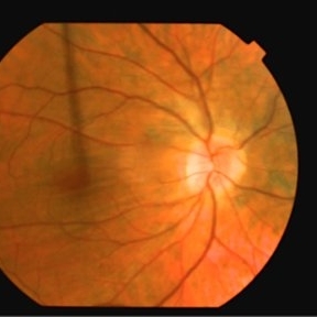
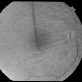
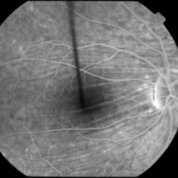
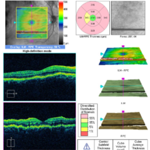
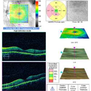
This 53 year old man was noted to have choroidal folds right greater than left. The visual acuity was normal at 20/15 bilaterally. The intraocular pressure was normal. The choroidal folds are visible on OCT, especially on the vertical cuts that image across the horizontal folds. Angiography revealed staining of the folds without CNVM, choroidal mass, or optic nerve edema. B scan did not reveal any retrobulbar masses. Choroidal folds can be due to hypotony, choroidal masses or retrobulbar masses, inflammatory conditions, choroidal neovascular membranes, optic nerve edema, scleral buckles, and idiopathic. In this case, they were thought to be idiopathic.
-
 Choroidal Folds
Nov 28 2014 by Thomas A. Ciulla, MD, MBA, FASRS
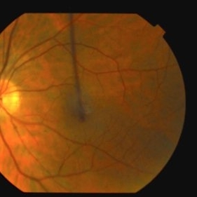
This 53-year-old man was noted to have choroidal folds right greater than left. The visual acuity was normal at 20/15. The choroidal folds are visible on OCT, especially on the vertical cuts that image across the horizontal folds. Angiography revealed staining of the folds without CNVM, choroidal mass, or optic nerve edema.
Photographer: Charlotte Harris
Condition/keywords: bilateral chorioretinal folds, choroidal folds
-
 Choroidal Folds
Nov 28 2014 by Thomas A. Ciulla, MD, MBA, FASRS
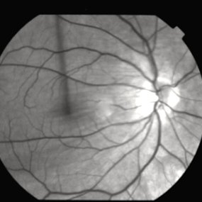
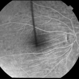
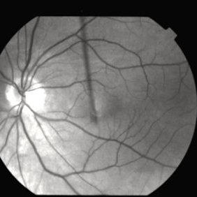
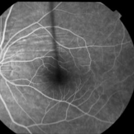
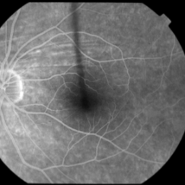
This 53 -year-old man was noted to have choroidal folds right greater than left. The visual acuity was normal at 20/15. The choroidal folds are visible on OCT, especially on the vertical cuts that image across the horizontal folds. Angiography revealed staining of the folds without CNVM, choroidal mass, or optic nerve edema.
Condition/keywords: bilateral chorioretinal folds, choroidal folds
-
 Choroidal Folds
Nov 28 2014 by Thomas A. Ciulla, MD, MBA, FASRS
This 53-year-old man was noted to have choroidal folds right greater than left. The visual acuity was normal at 20/15. The choroidal folds are visible on OCT, especially on the vertical cuts that image across the horizontal folds. Angiography revealed staining of the folds without CNVM, choroidal mass, or optic nerve edema.
Condition/keywords: bilateral chorioretinal folds, choroidal folds