 Initializing download.
-
 By Thomas A. Ciulla, MD, MBA, FASRS
Indiana University School of Medicine - Uploaded on Sep 15, 2014.
- Last modified by Caroline Bozell on Sep 16, 2014.
- Rating
- Appears in
- Choroidal Hemangioma
- Condition/keywords
- choroidal hemangioma, choroidal tumor, color fundus photograph
- Photographer
- Charlotte Harris
- Imaging device
- Fundus camera
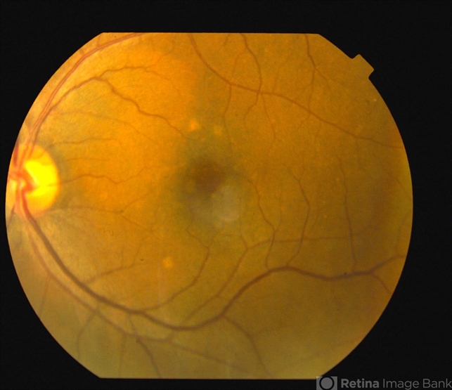
- Description
- Color fundus photography reveals a 2 x 3 DD well circumscribed orange-hued lesion superior nasal to the fovea. There were several small punched out spots inferior and superior to the fovea that were incidentally noted.


---thumb.jpg/image-square;max$79,0.ImageHandler "Metastatic Breast Cancer")

---thumb.jpg/image-square;max$79,0.ImageHandler "Choroidal Tumor, Breast Cancer")
---thumb.jpg/image-square;max$79,0.ImageHandler "Choroidal Tumor, Breast Cancer")


")