A 49 year old woman presented with metamorphopsia OS. She was noted to have a subtle orange-hued lesion superior nasal to the fovea. There were some small punched-out spots superior and inferior to the fovea, a common finding in the midwest, where POHS is prevalent. Fluorescein angiography revealed a well-circumscribed coarse hyperfluorescent vascular pattern within the lesion. There was no leakage during the late phase of the angiogram. OCT revealed subretinal fluid superior nasal to the fovea. B-scan showed a shallow echodense mass adjacent to the optic nerve with shallow overlying subretinal fluid. These findings were all characteristic of choroidal hemangioma.
-
 Choroidal Hemangioma
Sep 15 2014 by Thomas A. Ciulla, MD, MBA, FASRS
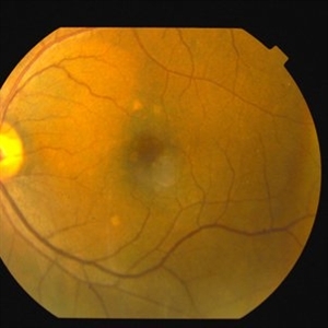
Color fundus photography reveals a 2 x 3 DD well circumscribed orange-hued lesion superior nasal to the fovea. There were several small punched out spots inferior and superior to the fovea that were incidentally noted.
Photographer: Charlotte Harris
Condition/keywords: choroidal hemangioma, choroidal tumor, color fundus photograph
-
 Choroidal Hemangioma
Sep 15 2014 by Thomas A. Ciulla, MD, MBA, FASRS
Red-free photography reveals a 2 x 3 DD well circumscribed lesion superior nasal to the fovea.
Photographer: Charlotte Harris
Condition/keywords: choroidal hemangioma, choroidal tumor, red-free
-
 Choroidal Hemangioma
Sep 15 2014 by Thomas A. Ciulla, MD, MBA, FASRS
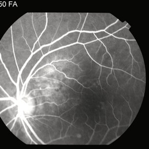
Mid-phase fluorescein angiography reveals a well-circumscribed coarse hyperfluorescent vascular pattern within the lesion.
Photographer: Charlotte Harris
Condition/keywords: choroidal hemangioma, choroidal tumor, FA mid phase
-
 Choroidal Hemangioma
Sep 15 2014 by Thomas A. Ciulla, MD, MBA, FASRS
Mid-phase fluorescein angiography reveals a well-circumscribed coarse hyperfluorescent vascular pattern within the lesion.
Photographer: Charlotte Harris
Condition/keywords: choroidal hemangioma, choroidal tumor, FA mid phase
-
 Choroidal Hemangioma
Sep 15 2014 by Thomas A. Ciulla, MD, MBA, FASRS
Mid-phase fluorescein angiography reveals a well-circumscribed coarse hyperfluorescent vascular pattern within the lesion.
Photographer: Charlotte Harris
Condition/keywords: choroidal hemangioma, choroidal tumor, FA mid phase
-
 Choroidal Hemangioma
Sep 15 2014 by Thomas A. Ciulla, MD, MBA, FASRS
Late phase fuorescein angiography reveals a well-circumscribed coarse hyperfluorescent vascular pattern within the lesion. There is no leakage.
Photographer: Charlotte Harris
Condition/keywords: choroidal hemangioma, choroidal tumor, late phase
-
 Choroidal Hemangioma
Sep 7 2014 by Thomas A. Ciulla, MD, MBA, FASRS
OCT macular thickness map reveals thickening superior nasal to the fovea.
Condition/keywords: choroidal hemangioma
-
 Choroidal Hemangioma
Sep 7 2014 by Thomas A. Ciulla, MD, MBA, FASRS
OCT shows subretinal fluid nasal to the fovea.
Condition/keywords: choroidal hemangioma
-
 Choroidal Hemangioma
Sep 7 2014 by Thomas A. Ciulla, MD, MBA, FASRS
B-scan shows shallow echodense lesion nasal to the optic nerve with shallow overlying subretinal fluid.
Condition/keywords: B scan ultrasound, choroidal hemangioma