-
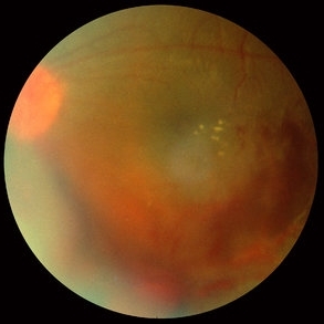 Sarcoidosis Panuveitis Slide 5
Oct 22 2012 by Ronald C. Gentile, MD
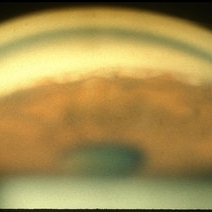
Fundus photo of the left eye reveal signs of vasculitis with exudate and a vitreous hemorrhage.
Photographer: The New York Eye & Ear Infirmary Department of Medical Imaging
Condition/keywords: sarcoidosis panuveitis, sarcoidosis vasculitis, vitreous hemorrhage
-
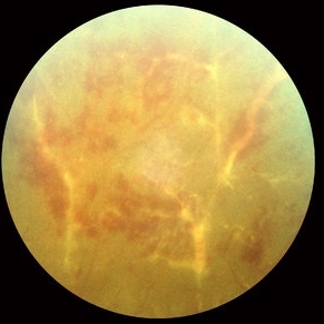 Sarcoidosis Panuveitis Slide 6
Oct 22 2012 by Ronald C. Gentile, MD
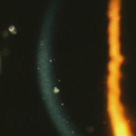
Fundus photo of the peripheral retina of the left eye reveal peri-vascular sheathing and ischemic vasculitis.
Photographer: The New York Eye & Ear Infirmary Department of Medical Imaging
Condition/keywords: sarcoidosis panuveitis, sarcoidosis vasculitis
-
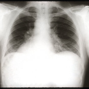 Sarcoidosis Panuveitis Slide 7
Oct 22 2012 by Ronald C. Gentile, MD
Chest x-ray revealed bihilar lymphadenopathy.
Photographer: The New York Eye & Ear Infirmary Department of Radiology
Condition/keywords: sarcoidosis panuveitis
-
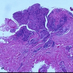 Sarcoidosis Panuveitis Slide 8
Oct 22 2012 by Ronald C. Gentile, MD
Biopsy of one of the sub-cutaneous nodules revealed non-caseating granulomas consistent with sarcoidosis.
Photographer: The New York Eye & Ear Infirmary Department of Pathology and Laboratory Medicine
Condition/keywords: sarcoidosis panuveitis
-
 Sarcoidosis Panuveitis Slide 3
Oct 22 2012 by Ronald C. Gentile, MD
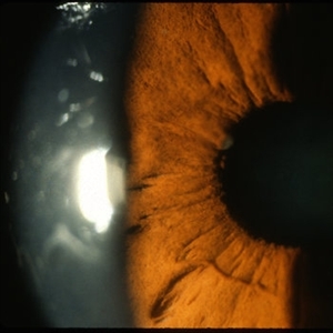
Gonioscopic photograph reveals peripheral anterior synechiae with granuloma involving the iris root and Bussaca nodules on the iris surface consistent with granulomatous uveitis.
Photographer: The New York Eye & Ear Infirmary Department of Medical Imaging
Condition/keywords: sarcoid granuloma, sarcoidosis panuveitis
-
 Sarcoidosis Panuveitis Slide 1
Oct 22 2012 by Ronald C. Gentile, MD
50-year-old African American women presented with decreasing vision in both eyes and floaters in the left eye. She had a history of asthma and sub-cutaneous skin nodules. Anterior segment examination reveal keratic precipitate on the corneal endothelium.
Photographer: The New York Eye & Ear Infirmary Department of Medical Imaging
Condition/keywords: sarcoidosis panuveitis
-
 Sarcoidosis Panuveitis Slide 4
Oct 22 2012 by Ronald C. Gentile, MD
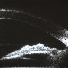
High frequency ultrasound biomicroscopy of the anterior chamber and angle images a granuloma involving the iris root and Bussaca nodules on the iris surface consistent with granulomatous uveitis.
Photographer: The New York Eye & Ear Infirmary Department of Medical Imaging
Condition/keywords: sarcoid granuloma, sarcoidosis panuveitis
-
 Sarcoidosis Panuveitis Slide 2
Oct 22 2012 by Ronald C. Gentile, MD
Anterior segment photo of the iris and pupillary margin shows a Koeppe nodule at the 9:30 position. Koeppe nodules consist of inflammatory cell precipitates.
Photographer: The New York Eye & Ear Infirmary Department of Medical Imaging
Condition/keywords: sarcoidosis panuveitis
A project from the American Society of Retina Specialists