-
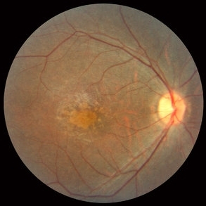 Cone Rod Dystrophy slide 1
Oct 22 2012 by Ronald C. Gentile, MD
30-year-old man with a history of early loss of color vision complained of recent nyctalopia and loss of peripheral vision in both eyes. ERG responses were reduced, greater on the photopic single-flash and flicker response compared to the scotopic ERG.
Photographer: The New York Eye & Ear Infirmary Department of Medical Imaging
Condition/keywords: cone dystrophy
-
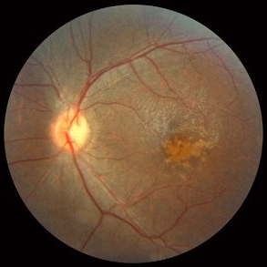 Cone Rod Dystrophy slide 2
Oct 22 2012 by Ronald C. Gentile, MD
Fundus examination revealed spotty pigmentary changes in the macular area with some peripheral depigmentation of the retinal pigment epithelium.
Photographer: The New York Eye & Ear Infirmary Department of Medical Imaging
Condition/keywords: cone dystrophy, retinal pigment epithelium
A project from the American Society of Retina Specialists