-
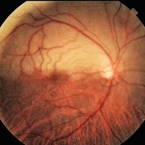 Branch Retinal Artery Oclussion
Mar 17 2024 by César Adrián Gomez Valdivia, MD
Decreased arterial blood flow to the retina leading to ischemic damage.
Photographer: Erika Paulina Ornelas Cazares
Imaging device: Topcon TRC-50DX
Condition/keywords: branch retinal artery occlusion (BRAO), oclussion
-
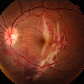 Choroidal Rupture
Sep 30 2025 by César Adrián Gomez Valdivia, MD
This fundus photograph shows curvilinear streaks of choroidal rupture radiating from the fovea, associated with subretinal hemorrhage. The rupture lines appear as crescent-shaped, whitish streaks representing a break in Bruch’s membrane, choriocapillaris, and retinal pigment epithelium.
Photographer: @eyemissu2
Imaging device: TOPCON TRX
Condition/keywords: Choroidal, Rupture
-
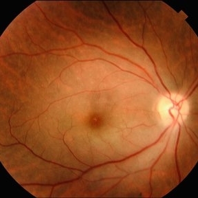 Central Retinal Artery Occlusion
Sep 30 2025 by César Adrián Gomez Valdivia, MD
This fundus photograph demonstrates the classic retinal whitening due to inner retinal ischemia, with a cherry-red spot at the fovea. The fovea appears red because it is nourished by the intact choroidal circulation, while the surrounding ischemic retina turns pale.
Photographer: @eyemissu2
Imaging device: TOPCON TRX
Condition/keywords: central retinal artery occlusion (CRAO)
-
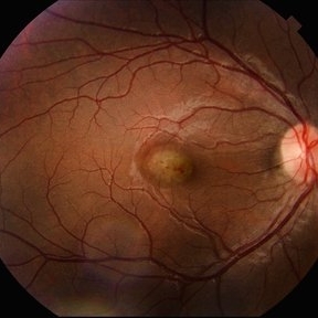 Idiopathic Choroidal Neovascularization
Sep 30 2025 by César Adrián Gomez Valdivia, MD
At the foveal area, there is a yellowish-greenish elevated lesion with indistinct borders, corresponding to a subfoveal choroidal neovascular membrane (CNV). There are subtle overlying changes including mild retinal pigment epithelium (RPE) disruption, and small hemorrhagic spots suggesting active leakage. Surrounding the lesion, there are faint retinal folds or striae, likely due to localized subretinal fibrosis or traction.
Photographer: @eyemissu2
Imaging device: TOPCON TRX
Condition/keywords: Idiopathic Choroidal Neovascularization
-
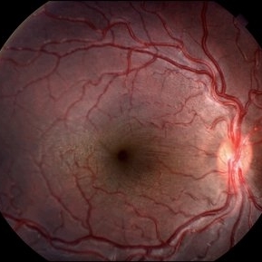 Congenital Vascular Tortuosity
Sep 30 2025 by César Adrián Gomez Valdivia, MD
The retinal vasculature is remarkable for pronounced vascular tortuosity, more evident in the superior and inferior temporal arcades. Both arteries and veins follow exaggerated curves and bends, deviating from the expected smooth course across the retina.
Photographer: @eyemissu2
Imaging device: TOPCON TRX
Condition/keywords: Congenital, Tortuosity, vascular tortousity
A project from the American Society of Retina Specialists