-
 Congenital Retinal Vessel Tortuosity
Apr 2 2024 by Pablo Angel Garcia Uribe
Fundus photograph of a 29-year-old man with bilateral congenital retinal vessel tortuosity. This image shows the sinuous course of retinal arterioles and a shiny internal limiting membrane.
Photographer: Pablo Ángel García-Uribe, Clínica Oftalmológica Salauno, Mexico City
Imaging device: NIDEK OCT RS-330 Duo 2
Condition/keywords: abnormal retinal vessel, anomalous vessels, Retina, tortuous vessels
-
 Persistent Hyperplastic Primary Vitreous Associated With Retrolental Fibrovascular Membrane
Aug 8 2025 by Pablo Angel Garcia Uribe
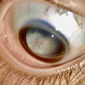
A 12-year-old Mexican male, asymptomatic, referred for evaluation after incidental finding of partial leukocoria on routine ophthalmologic examination. Slit-lamp evaluation revealed a fibrovascular retrolental membrane without evidence of retinal traction, associated with a fibrous stalk connecting to the optic disc. The stalk showed near-complete involution, consistent with a remnant of persistent fetal vasculature (posterior type).
Photographer: Pablo Angel García-Uribe, Clínica Oftalmológica Salauno, Mexico City
Condition/keywords: Persistent Hyperplastic Primary Vitreous Fibrovascular membrane
-
 Papillophlebitis Salauno
Sep 3 2025 by Pablo Angel Garcia Uribe
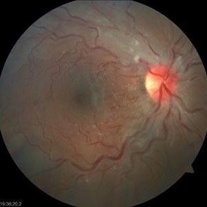
Fundus photograph of a 24-year-old woman, previously healthy, with a history of recreational inhaled cannabis use, presented with a 24-hour history of photopsias and mild decrease in visual acuity, associated with a subtle relative central scotoma in the right eye. On ophthalmic examination, the anterior segment of both eyes was unremarkable. Best-corrected visual acuity was slightly reduced in the right eye and normal in the left. Fundus biomicroscopy of the right eye revealed moderate disc edema with hyperemia and well-defined margins, accompanied by venous engorgement and tortuosity, predominantly affecting the venules. No retinal hemorrhages were observed. Additionally, retinal thickening was noted along the temporal arcades, with apparent foveal sparing. The left eye showed no pathological findings. Based on the patient’s age, the acute onset of symptoms, the fundoscopic features, and the absence of systemic risk factors, the clinical presentation was consistent with papillophlebitis.
Photographer: Clínica Oftalmológica Salauno
Imaging device: Visucam 524, Carl Zeiss Meditec AG, Jena, Germany
Condition/keywords: papillophlebitis
-
 Fluorescein Angiography Papillophlebitis Salauno
Sep 3 2025 by Pablo Angel Garcia Uribe
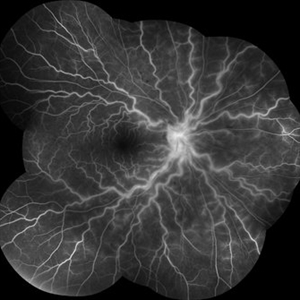
In the arteriovenous phase, fluorescein angiography demonstrated venous engorgement and tortuosity, with relative incompetence of the venous walls leading to mild leakage. Optic disc staining with late leakage was also observed. There was no evidence of significant capillary non-perfusion, and only subtle perivenous leakage was noted. The foveal region remained spared.
Photographer: Optom. Marilyn Alvarez Monroy, Clínica Oftalmológica Salauno
Imaging device: Visucam 524, Carl Zeiss Meditec AG, Jena, Germany
Condition/keywords: FA late phase leakage, retina
A project from the American Society of Retina Specialists