-
 Retinal Detachment Secondary to Anomalous PVD
Mar 13 2025 by Fabricio Dolores
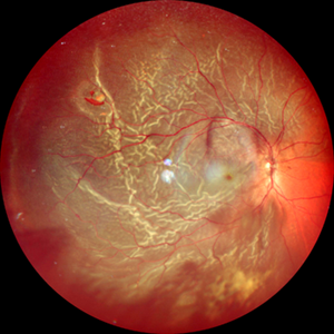
This color wide-field clinical image depicts the right eye of a female patient who experienced a sudden loss of vision one month earlier. She was initially diagnosed with a vitreous hemorrhage and managed with conservative treatment. Upon presentation to our institute one month later, a superior rhegmatogenous retinal detachment was identified, extending across the 12 o’clock meridian. This was accompanied by an inferior vitreous hemorrhage and a solitary superior retinal lesion located at M11 in the superior triangle of the ora serrata, in alignment with Lincoff's second law.
Photographer: Fabricio Dolores-Villanueva, MD
Imaging device: Nidek Mirante
Condition/keywords: Retinal Detachment
A project from the American Society of Retina Specialists