-
 Choroidal Detachment
Jan 17 2022 by Logan ryzenga
Left ultra-wide field photograph of an 81-year old female with a choroidal detachment affecting her left eye. Patient had a stent placed November, 2021 and following the procedure she complains of variable blurred vision and severe constricted visual fields. She presented at our office with flashes a month prior but without pain or floaters.
Photographer: Logan Ryzenga
Imaging device: Optos California
Condition/keywords: choroidal detachment, fundus photograph, left eye, Optos, pseudocolor, superior retina, ultra-wide field imaging
-
 Branch Retinal Vein Occlusion with Multifactorial Macular Edema and Epiretinal Membrane
Oct 3 2024 by Logan ryzenga
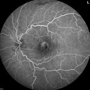
Fluorescein angiogram of a 62 year old woman with cystoid macular edema from concurrent Epiretinal Membrane and Branch Retinal Vein occlusion. She has an extensive history of anti-VEGF injections with stable but unresolved macular edema. Following angiography, it was determined that an epiretinal membrane peel would be indicated in an attempt to achieve resolution of macular edema.
Photographer: Logan Ryzenga
Imaging device: Heidelberg Spectralis
Condition/keywords: 55-degrees, branch retinal vein occlusion (BRVO), cystoid macular edema (CME), epiretinal membrane (ERM), Fluorescein angiography, heidelberg spectralis, hyperfluorescence, leakage, left eye, OS, wide angle imaging
-
 Occlusive Retinal Vasculitis
Oct 3 2024 by Logan ryzenga
4 view ultra-widefield Optos fluorescein angiogram in the left eye of a 39 year old woman occlusive retinal vasculitis with four quadrant Kyrieleis plaques. This is a showcase of a suspected, rarely reported, and atypical presentation of Behcet's Syndrome.
Photographer: Logan Ryzenga
Imaging device: Optos California
Condition/keywords: Behcet's Disease, Behcet's uveitis, Fluorescein angiography, fluorescein leakage, kyrieleis plaques, non-perfusion, OPTOS, OPTOS CALIFORNIA, ultra-wide field imaging, Uveitis
A project from the American Society of Retina Specialists