 Initializing download.
-
 By Isaac Agranoff
Retina and Uveitis Center
Co-author(s): Muge Kesen, MD - Uploaded on Mar 21, 2024.
- Last modified by Joshua Friedman on Mar 22, 2024.
- Rating
- Appears in
- Miscellaneous
- Condition/keywords
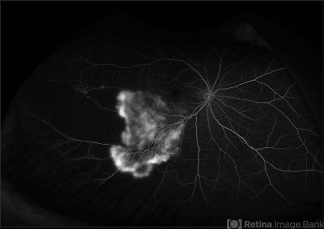
- branch retinal vein occlusion (BRVO), Neovascularisation elsewhere (NVE), Optos, EYLEA
- Photographer
- Isaac Agranoff
- Imaging device
-
Scanning laser ophthalmoscope
Optos California - Description
- Fundus angiography photograph of a 63 year old male presenting with worsening blurry vision OD for 4 years with new transient floaters (vision 20/160 PH 20/60). Fluorescein angiography revealed significant capillary non-perfusion corresponding to the area, with peripheral vascular remodeling. Physician recommended anti-VEGF therapy and FA-guided supplemental PRP given the size of the NVE.


 Caused due Branch Retinal Vein Occlusion (BRVO)")
 after Anti VEGF Treatment")
")


