 Initializing download.
-
 By MEENAL SONI
ASG Eye Hospital Jodhpur
Co-author(s): Dr. Pravin Jain; Consultant and Head, Dr. Pramod K Suman; Consultant, VR services ASG eye Hospital, Jodhpur - Uploaded on Jan 7, 2024.
- Last modified by Joshua Friedman on Jan 8, 2024.
- Rating
- Appears in
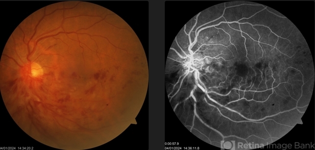
- Inferior HRVO
- Condition/keywords
- HRVO, cystoid macular edema (CME)
- Photographer
- Dr. Meenal Soni, Fellow VR, ASG eye Hospital Jodhpur
- Imaging device
-
Fundus camera
Visucam - Description
- A middle aged man presented to our OPD with sudden DOV in LE since 2 days. There was no history of any systemic illness. On fundus examination of LE we found dilated tortuous veins on inferior half of retina and multiple superficial retinal haemorrhages with corresponding disc margin blurring, elevation and macular oedema suggestive of inferior hemi-retinal vein occlusion with macular oedema. On fluorescein angiography there was corresponding blocked fluorescence, dilated tortuous veins and capillary non perfusion areas.
")
")
")
")
")
")


---thumb.jpg/image-square;max$79,0.ImageHandler "Intermediate Uveiris and CME")
---thumb.jpg/image-square;max$79,0.ImageHandler "Intermediate Uveitis and CME")