 Initializing download.
-
---thumb.jpg/image-square;max$48,0.ImageHandler) By Susanna S. Park, MD, PhD
By Susanna S. Park, MD, PhD
University of California Davis Eye Center - Uploaded on Jan 10, 2014.
- Last modified by Susanna S. Park, MD, PhD on Jul 9, 2014.
- Rating
- Appears in
- Surgical
- Condition/keywords
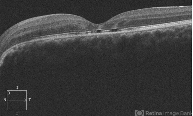
- optical coherence tomography (OCT), maculopathy, macular schisis
- Photographer
- Ellen Redenbo, University of California Davis Eye Center
- Imaging device
- Optical coherence tomography system
- Description
- OCT image taken 1 year after vitrectomy with gas tamponade for macular schisis and detachment and outer lamellar hole associated with optic pit shows normal macular morphology with only mild disruption of the foveal photoreceptor layer.

")


")
")
")


