 Initializing download.
-
 By Jessica Hampton, BS
Stanford Medical School
Co-author(s): Dr. Diana Do, Stanford Medicine, Byers Eye Institute - Uploaded on Oct 25, 2023.
- Last modified by Joshua Friedman on Oct 25, 2023.
- Rating
- Appears in
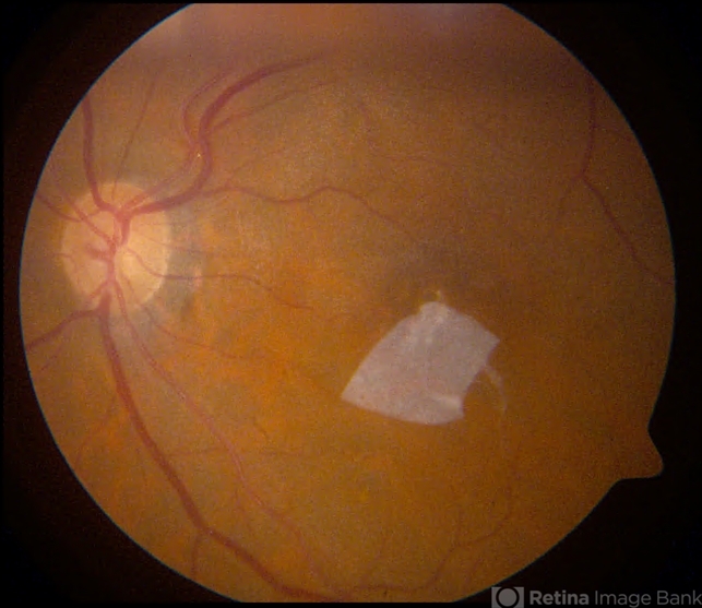
- Chronic Full Thickness Macular Hole
- Condition/keywords
- full thickness macular hole, amniotic membrane graft, fundus photograph
- Photographer
- Dr. Diana Do, Stanford Medicine, Byers Eye Institute
- Imaging device
- Fundus camera
- Description
- Fundus photograph of a 67-year-old woman with a history of recurrent, chronic full-thickness macular hole in the left eye repaired with an amniotic membrane graft, seen at 2 years follow-up.
")
")
")


---thumb.JPG/image-square;max$79,0.ImageHandler "Welding arc maculopathy")


---thumb.jpg/image-square;max$79,0.ImageHandler "Normal Fundus Photo")