Search results (0 results)
-
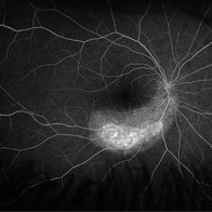
 Lupus Vasculitis
Feb 13 2013 by From the Collections of Thomas M. Aaberg, MD and Thomas M. Aaberg Jr., MD
Obliterative peripheral vasculitis.
Condition/keywords: ischemia, lupus
-
---thumb.jpg/image-square;max$300,300.ImageHandler) Lupus Vasculitis Angiogram
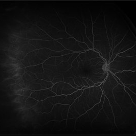
Lupus Vasculitis Angiogram
Feb 13 2013 by From the Collections of Thomas M. Aaberg, MD and Thomas M. Aaberg Jr., MD
FA, lupus vasculitis angiogram.
Condition/keywords: lupus, obliterative peripheral vasculitis, retinal ischemia
-
 Behcet's Syndrome
Apr 1 2016 by Nichole Lewis
Behcet's Syndrome
Photographer: Nichole Lewis - Pennsylvania Retina Specialists, Camp Hill, PA
Condition/keywords: Behcet's Disease, obliterative peripheral vasculitis
-
 Behcet's Syndrome
Apr 1 2016 by Nichole Lewis
Behcet's Syndrome
Photographer: Nichole Lewis - Pennsylvania Retina Specialists, Camp Hill, PA
Condition/keywords: Behcet's Disease, obliterative peripheral vasculitis
-
 Xenon Photocoagulation Marks in Vasculitis
Jan 10 2017 by Manish Nagpal, MD, FRCS (UK), FASRS
Patient gave history of being treated 25 years back with xenon photocoagulation for peripheral vasculitis.
Photographer: Pooja Barot
Condition/keywords: vasculitis, xenon photocoagulation
-
 Acute Zonal Occult Outer Retinopathy (AZOOR) FA, Fluorescein Angiography, Peripheral Vasculitis
Jan 19 2022 by James B. Soque, CRA, OCT-C, COA, FOPS
Acute Zonal Occult Outer Retinopathy (AZOOR). Peripheral Vasculitis OD. Fluorescein angiography showing vasculitis in the far right periphery 8-10 o'clock. 46-year-old white male, VA CC 20/16, 20/12.5, has had recurrent vasculitis for 11 years. No treatment.
Photographer: James Soque, CRA, OCT-C, COA, FOPS, Island Retina, Shirley, NY
Imaging device: Optos California
Condition/keywords: acute zonal occult outer retinopathy (AZOOR), FA early phase, fluorescein angiogram (FA), Peripheral Vasculitis, ultra-wide field imaging
-
 Peripheral Retinal Vasculitis
May 27 2020 by Olivia Rainey
Ultra-widefield fluorescein angiogram of a 58-year-old female with possible peripheral vasculitis. There was no venous access for this patient, so the fluorescein was administered orally. The image was taken at 7:33 after oral administration. The physician stated that the peripheral nonperfusion could be a sign of previous vasculitis, although could also be a result of uncontrolled diabetes. She was asked to obtain additional bloodwork in order to rule out sarcoidosis, as well as sickle cell. It does not appear the nonperfusion has progressed since her last evaluation. Her vision was 20/40 in the right eye at the time the image was taken.
Photographer: Olivia Rainey, OCT-C, COA
Imaging device: Optos California
Condition/keywords: diabetes, fluorescein angiogram (FA), hypertensive retinopathy, non-perfusion, Optos, oral fluorescein, peripheral retinal vasculitis, ultra-wide field imaging
-
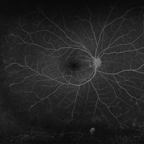 Obliterative Peripheral Vasculitis
Apr 3 2019 by HECTOR LUIS VILLARROEL GUIZAR, MD, RETINA FELLOW
Ultra-wide angle retinal angiography of an 27-year-old with retinal vasculitis with late stain of the vascular wall and obliteration of the peripheral vasculature.
Photographer: HECTOR VILLARROEL, HOSPITAL DE LA LUZ, MEXICO CITY
Imaging device: OPTOS
Condition/keywords: obliterative peripheral vasculitis
-
 Atypical Tubercular Occlusive Peripheral Retinal Vasculitis
Jun 21 2024 by Tejaswita Verma
Follow up right eye fundus photograph of a 27 year old male with vision 6/12 , diagnosed with systemic tuberculosis(mediastinal lymphadenopathy on chest CT) on oral steroids, and started on ATT .We can see a parafoveal sub-ILM hemorrhage with vascular sheathing and hemorrhages in inferior and temporal quadrants . The patient was advised anti-VEGF intravitreal injection, later sectoral laser after resolution of inflammation
Photographer: DR. TEJASWITA VERMA
Imaging device: MIRANTE
Condition/keywords: obliterative peripheral vasculitis, ocular tuberculosis
-
 FFA in Atypical Tubercular Peripheral Occlusive Retinal Vasculitis
Jun 21 2024 by Tejaswita Verma
Right eye FFA montage of a 27 year male with peripheral occlusive tubercular vasculitis, showing CNP areas inferiorly and temporally, leakages and blocked fluorescence due to hemorrhages. The patient was advised intravitreal anti-VEGF injection and later sectoral laser once inflammation subsides.
Photographer: DR. TEJASWITA VERMA
Imaging device: MIRANTE
Condition/keywords: obliterative peripheral vasculitis, ocular tuberculosis
 Loading…