Search results (14 results)
-
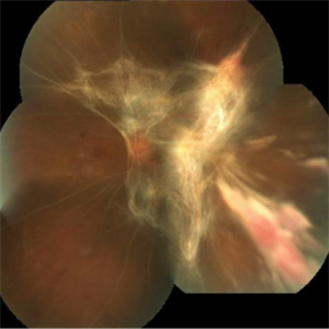
 Post Combined Surgery of Cataract, TRD & Vitreous Hemorrhage
Jun 27 2024 by Sanauddin Samejo , Diploma (Ophthalmic Technician Training Course)
A 27 year-old diabetic female visited the clinic one week after combined surgery of cataract, tractional retinal detachment and vitreous hemorrhage.
Photographer: Sanauddin Samejo, Burjeel Hospital, Abu Dhabi, UAE
Imaging device: Silver Stone Optos
Condition/keywords: Combined Surgery Cataract Tractional Retinal Detachment Vitreous Hemorrhage, POST SURGERY, Retinal Detachment, TRD
-
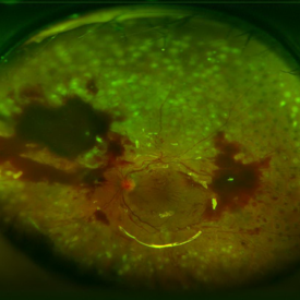
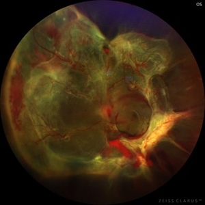 Diabetic traction retinal detachment
Jan 9 2023 by JORGE SOBERANES
Proliferative diabetic retinopathy with extensive traction retinal detachment in a patient with type 1 diabetes mellitus.
Photographer: Dr. Jorge I. Soberanes, Asociación para Evitar la Ceguera en México.
Imaging device: Zeiss Clarus 700
Condition/keywords: Retinal Detachment, tractional retinal detachment
-
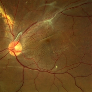 Displaced & folded macula
Oct 10 2022 by Ricardo Leitão Guerra
Tractional retinal detachment due to sickle cell retinopathy leading to a displaced and folded appearance of the macula in this 36-yo male. Subretinal bands are also noticed crossing the macula towards inferior retinal detachment area.
Photographer: Ricardo Leitão Guerra
Imaging device: Clarus 700 - Zeiss
Condition/keywords: folds, sickle cell retinopathy, subretinal bands, tractional retinal detachment
-
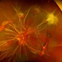 Sickle Cell Retinopathy
Nov 5 2022 by Mateus Queiroz Corrêa, MD
19 -year-old young man with combined rhegmatogenous and tractional retinal detachment secondary to a proliferative sickle retinopathy ( stage V)
Photographer: Mateus Corrêa, Sorocaba Eye Bank Hospital
Imaging device: Optos California
Condition/keywords: Retinal detachment, sickle cell retinopathy
-
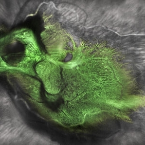 Green Goblin Detachment
Jan 13 2022 by Netan Choudhry, MD, FRCS(C) FASRS
Tractional retinal detachment with macular hole in a 76-year-old female.
Photographer: John Golding BA, Vitreous Retina Macula Specialists of Toronto, OCTane Imaging Lab
Imaging device: Multicolor fundus photo taken on the Spectralis OCT2 (Heidelberg Engineering GmbH).
Condition/keywords: macular hole, Multispectral imaging, tractional retinal detachment
-
 Proliferative Diabetic Retinopathy with Traction Retinal Detachment OS
Apr 28 2020 by Pauline T Merrill, MD, FASRS
Fundus photographs of an 29-year-old man with PDR, TRD, VH OS.
Photographer: Karen Parque, Illinois Retina Associates
Condition/keywords: proliferative diabetic retinopathy (PDR), tractional retinal detachment
-
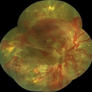
 Diabetic Macular TRD
Jan 10 2020 by Somnath Chakraborty, MD
Fundus Montage image of the left eye of a 48-year-old type 2 diabetic with post PRP macular extensive tractional retinal detachment involving macula.
Photographer: Pulak Roy
Condition/keywords: diabetic retinopathy, proliferative diabetic retinopathy (PDR), tractional retinal detachment, vitrectomy, vitreomacular surgery
-
 Proliferative Diabetic Retinopathy (PDR)
Jul 4 2018 by Deepak Bhojwani, MS
Colour Fundus Photograph of a 66-year-old diabetic male with large fibro-vascular proliferative vessels causing subhayolid haemorrhage and tractional retinal detachment involving posterior pole.
Photographer: Deepak Bhojwani
Condition/keywords: diabetes, neovascularization (NV), subhyaloid hemorrhage, tractional retinal detachment
-
 Extensive Tractional Retinal Detachment in Proliferative Diabetic Retinopathy
Jun 4 2018 by Diva Kant Misra, MBBS, DO, DNB, MNAMS, FVRS
Montage fundus photograph of a 54-year-old male diabetic patient showing extensive TRD with PDR.
Photographer: DIVA KANT MISRA
Condition/keywords: diabetes, proliferative diabetic retinopathy (PDR), tractional retinal detachment
-
 TRD After Surgical Repair
Sep 14 2017 by Theodore Leng, MD, MS, FASRS
TRD after surgical repair.
Condition/keywords: proliferative diabetic retinopathy (PDR), tractional retinal detachment
-
 Tractional Retinal Detachment
Jul 29 2017 by FELIPE PEREIRA
Fundus photograph of an 40-year-old woman with diabetes mellitus diagnosed 20 years ago in insulin use. This image is from her right eye and it was diagnosed with severe total tractional retinal detachment. The best correct visual acuity was hand motion in this eye.
Photographer: Felipe Pereira, Federal University of Sao Paulo, Sao Paulo, Brazil
Imaging device: VISUCAM 524 Fundus Imaging
Condition/keywords: diabetes, tractional retinal detachment
-
 TRD Secondary to PDR
Feb 10 2015 by Matt Poe, COA
This patient has a long standing tractional detachment secondary to his proliferative diabetic retinopathy. Patient is CF in this eye.
Photographer: Matt Poe, COA. Northwest Arkansas Retina Associates, Springdale, AR.
Imaging device: Heidelberg HRA
Condition/keywords: diabetes, proliferative diabetic retinopathy (PDR), red-free, tractional retinal detachment
-
000---thumb.jpg/image-square;max$300,300.ImageHandler) Fundus Panorama Finding of Tractional Retinal Detachment Due to Proliferative Diabetic Retinopathy
Fundus Panorama Finding of Tractional Retinal Detachment Due to Proliferative Diabetic Retinopathy
Dec 25 2013 by Dong Yoon Kim, MD
47-year-old woman visited our clinic for decreased visual acuity on her right eye. Her visual acuity of right eye was hand motion. Fundus examination showed traction retinal detachment.
Condition/keywords: fundus photograph, tractional retinal detachment
-
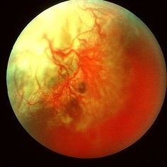 Tractional Retinal Detachment Due To Proliferative Diabetic Retinopathy
May 8 2013 by Jerald A. Bovino, MD
Fundus photograph of a tractional retinal detachment due to proliferative diabetic retinopathy.
Condition/keywords: tractional retinal detachment
 Loading…