Search results (5 results)
-
 Pericentral Retinitis Pigmentosa
Sep 6 2024 by Mauricio Bayram-Suverza, MD
A 65-year-old male patient reports experiencing bilateral blind spots that have gradually intensified over time. Genetic testing was unrevealing. The fundus autofluorescence image shows a hypoautofluorescent ring in the posterior pole, especially nasal to the nerve and along arcades.
Photographer: Mauricio Bayram-Suverza, Casey Eye Institute, OHSU.
Imaging device: Optos California
Condition/keywords: fundus autofluorescence (FAF), inherited retinal disease, nyctalopia, retinal dystrophy, retinitis pigmentosa
-
 Rod Cone dystrophy
Nov 29 2022 by Niloofar Piri, MD
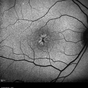
Fundus autofluorescence of the left eye in a 58 yo male with rod cone dystrophy. He presented with night blindness and peripheral vision loss since youth and recent decrease in central vision for the past 10 years. Notice multiple coin shaped hypoautofluorescent pacthes within central 20 degrees which are coalescing centrally. (fundus photo uploaded separately) He has one pathogenic variants of both CEP290 and PRPH2 genes.
Photographer: Sean Kelso, Saint Louis University
Condition/keywords: hereditary retinal degeneration, hereditary retinal dystrophy, rod cone dystrophy
-
 Rod cone dystrophy autofluorescence
Sep 19 2022 by Kenneth Fong
34 year old male with colour blindness and loss of visual field
Condition/keywords: retinal dystrophy
-
 Gyrate Atrophy
Jan 6 2019 by Hashim Ali Khan, OD, FAAO
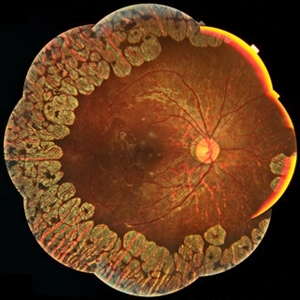
Montage of Multiple Fundus Photographs from the right eye of a 25-year-old woman with gyrate atrophy.
Photographer: Ahmed Abbass
Imaging device: Topcon TRC-NW8F
Condition/keywords: gyrate atrophy, hereditary retinal dystrophy, retinal dystrophy
-
 Retinal Dystrophy of 24-Year-Old Male/ AF OD
Nov 25 2015 by Zach Dupureur
Fluorescein angiography of a 24-year-old male. Juvenile retinoschisis on OCT. FA shows outer retinal staining. Could be associated with Goldman Farve Syndrome.
Photographer: Zach Dupureur OCT-C
Imaging device: Heidelberg Spectralis
Condition/keywords: Goldmann-Favre Syndrome, juvenile retinoschisis, retinal dystrophy
 Loading…