Search results (5 results)
-
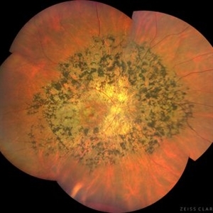
 Rod Cone dystrophy
Nov 29 2022 by Niloofar Piri, MD
Fundus autofluorescence of the left eye in a 58 yo male with rod cone dystrophy. He presented with night blindness and peripheral vision loss since youth and recent decrease in central vision for the past 10 years. Notice multiple coin shaped hypoautofluorescent pacthes within central 20 degrees which are coalescing centrally. (fundus photo uploaded separately) He has one pathogenic variants of both CEP290 and PRPH2 genes.
Photographer: Sean Kelso, Saint Louis University
Condition/keywords: hereditary retinal degeneration, hereditary retinal dystrophy, rod cone dystrophy
-
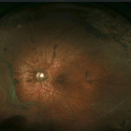
 Tapetoretinal Degeneration
Sep 7 2022 by JEFFERSON R SOUSA, Tecg.º (Biomedical Systems Technology)
Patient 52 years old, Male, progressive loss of vision since the age of 20. Retinography showed mobilization of pigments in osteoblasts, extensive area of atrophy of the pigmentary epithelium and choroid. On fluorescein angiography, typical changes following the characteristic patterns of paracentra retinal retinitis pigmentosa. Autofluorescent fundus with a sectorial autohypofluorescence pattern in the regions of atrophies.
Photographer: JEFFERSON ROCHA DE SOUSA - Retinal Department at Instituto Dr. Suel Abujamra Sao Paulo-Brazil
Imaging device: Clarus 700 - Zeiss, composite of four 135 degree images.
Condition/keywords: pericentral retinitis pigmentosa, tapeoretinal degeneration
-
 Macula-Sparing GRT RRD
Jul 6 2017 by Andrew A. Moshfeghi, MD, MBA, FASRS
Wide-field fundus photograph of a 43-year-old myopic man with a history of lattice retinal degeneration status posterior barrier laser performed elsewhere who presented with a giant-retinal tear associated retinal detachment of the right eye.
Photographer: Jay Jiang, University of Southern California Roski Eye Institute
Imaging device: Optos California
Condition/keywords: acute retinal detachment, giant retinal tear, lattice degeneration
-
 Stickler Syndrome
Dec 8 2016 by Aleksandra V. Rachitskaya, MD, FASRS
Optos wide-field fundus image of a patient with Stickler Syndrome and COL2A1 gene mutation. Patient has perviously undergone prophylactic laser. Lattice, vitreous veils, and laser scars are seen.
Photographer: Anne Pinter, Cole Eye Institute, Cleveland Clinic
Condition/keywords: Stickler Syndrome, vitreoretinal degeneration
-
 ARMD With Geographic Atrophy, Peripheral Degeneration
Dec 6 2013 by James B. Soque, CRA, OCT-C, COA, FOPS
92-year-old white female with exudative macular degeneration, geographic atrophy, and peripheral retinal degeneration.
Photographer: James Soque, CRA COA, Island Retina, Shirley, New York
Imaging device: Topcon TRC 50DX with OIS 10.6.45
Condition/keywords: fundus photograph, geographic atrophy
 Loading…