Search results (3 results)
-
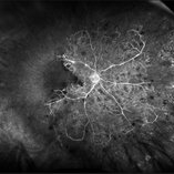
 High risk Proliferative Diabetic Retinopathy treated with Pan Retinal Photocoagulation
Nov 5 2022 by Somnath Chakraborty, MD
A Fundus Photo Montage of 43 year old Asian Male with Type 2 Diabetes Mellitus since 7 years who presented with sudden onset diminition of vision in his Left eye. BCVA OS was 20/200. He was diagnosed to have Pre retinal bleed due to Proliferative Diabetic Retinopathy and was treated with Pan Retinal Photocoagulation. This image shows a large neo-cascular frond at the disc and superior to it with Pre-retinal bleed and Fresh laser marks along
Photographer: Pulak Roy
Condition/keywords: diabetic blindness, diabetic retinopathy vitrectomy study (DRVS), fresh laser burns, laser photocoagulation, preretinal hemorrhage, proliferative diabetic retinopathy (PDR)
-
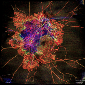 Flame of the Forest
Apr 9 2020 by Daraius N Shroff, MS FMRF FRCS
A 54-year-old man with DM for 15 years. The left eye had a visual acuity of 20/40. Wide field swept source OCTA revealed branching out central neovascular trunk vessels from the disc with terminal loops, along with exuberant proliferation of irregular small-calibre fine new vessels. The patient underwent OCTA guided pan retinal photocoagulation.
Photographer: Anuj Choudhary, Shroff Eye Centre, New Delhi
Imaging device: Zeiss Plex Elite 9000
Condition/keywords: proliferative diabetic retinopathy (PDR)
-
 Ischemic Central Retinal Vein Occlusion
Jan 24 2019 by Nichole Lewis
76-year-old woman with an ischemic central retinal vein occlusion, severely attenuated and sclerotic vessels and scattered retinal hemorrhages. Vision decrease over 1 year. VA 20/CF. Patient is returning for pan retinal photocoagulation.
Photographer: Nichole Lewis
Imaging device: Optos
Condition/keywords: attenuated vessels, central retinal vein occlusion (CRVO), hemorrhage, ischemic CRVO, sclerotic vessels
 Loading…