Search results (2 results)
-
 SLE Retinopathy
Nov 14 2016 by Mitzy E Torres Soriano, MD
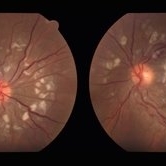
25-year-old female patient with systemic lupus erythematosus. Photographs show cotton wool spots, intraretinal hemorrhages and vascular tortuosity. FA demonstrated retinal vasculitis and OCT revealed cystoid macular edema. In this case diagnosis of SLE was made after ocular manifestation.
Photographer: Grupo Laser Vision, Rosario, Argentina
Condition/keywords: cotton wool spots, occlusive retinal vasculitis, occlusive vasculitis, systemic lupus erythematosus, vasculopathy
-
 Subhyaloid Hemorrhage
Jul 7 2015 by Hamid Ahmadieh, MD
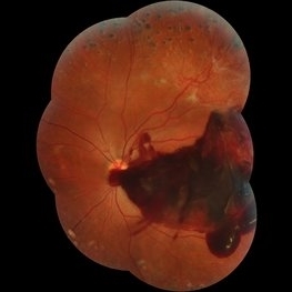
Color fundus photograph of the left eye of a 25-year-old woman with severe subhyaloid hemorrhage due to an advanced vasoproliferative vitreoretinopathy secondary to a severe idiopathic occlusive retinal vasculitis.
Photographer: Soulmaz Shahmohammad, Negah Eye Center, Tehran, Iran
Condition/keywords: color fundus photograph, occlusive vasculitis, subhyaloid hemorrhage
 Loading…