Search results (6 results)
-
 Retinoschisis
Mar 28 2021 by JEFFERSON R SOUSA, Tecg.º (Biomedical Systems Technology)
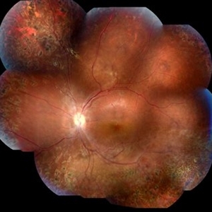
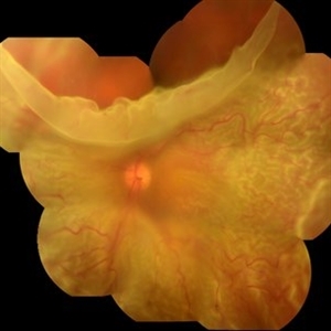
A 14-year-old male patient was admitted for visual evaluation. Visual acuity s/c in the right eye and 20/80 in the left eye. According to family members, he reported low vision since childhood. He had already undergone treatment with photocoagulation in another service to which he had a diagnostic hypothesis of Coats' disease. Laboratory tests were requested (HIV, TOXO, TOXOCARIASIS, ECA, VDRL, PPD). In the evaluation it was observed important exudation in the posterior pole, some vascular irregularities in the right eye. In the left eye, there is retinoschisis affecting the entire posterior pole and the region nasal to the optic disc, macula with a characteristic aspect of a cartwheel. Well exemplified by OCT-A (Structrure Deep: IPL - 25, OPL - 25).
Photographer: JEFFERSON R SOUSA - Study Center and Ophthalmological Research Dr. Andre M V Gomes, Institute Dr. Suel Abujamra São Paulo-Brazil
Imaging device: Topcon TRC-50 DX, Imaginet 4.0, angle de 50 graus. Flash 50w-s
Condition/keywords: Coats' disease, retinoschisis
-
 Gyrate Atrophy
Oct 30 2020 by JEFFERSON R SOUSA, Tecg.º (Biomedical Systems Technology)
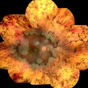
Female patient, 28-year-old, with low vision in both eyes since childhood. In routine examination, important changes were observed with atrophic, symmetrical and bilateral aspects with apparently preservation of the central retina.
Condition/keywords: gyrate atrophy
-
 Uveitis Posterior
Jul 19 2019 by JEFFERSON R SOUSA, Tecg.º (Biomedical Systems Technology)
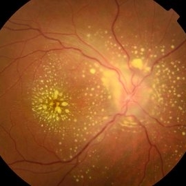
A 23-year-old male patient attended the clinic with low vision of the right eye. In the evaluation it presented important fundoscopical alterations like retinal exudations in the posterior pole and nasal retina, aspects of macular star. It was proven that it was a posterior uveitis.
Photographer: JEFFERSON R SOUSA - Study Center and Ophthalmological Research Dr. Andre M V Gomes, Institute Dr. Suel Abujamra São Paulo-Brazil
Imaging device: Topcon TRC-50 DX, Imaginet 4.0, angle de 50 graus. Flash 50w-s
Condition/keywords: uveitis
-
 Retinal Detachment
Feb 17 2018 by JEFFERSON R SOUSA, Tecg.º (Biomedical Systems Technology)
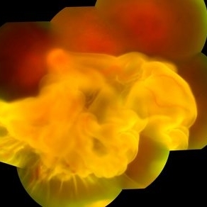
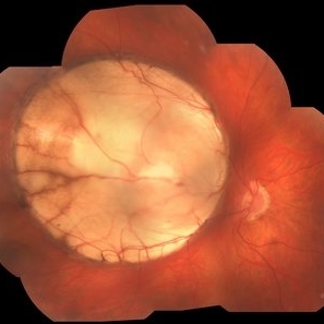
A 42-year-old patient complained of low vision in the left eye. In retinal mapping and background color photography, extensive retinal detachment was observed.
Photographer: JEFFERSON R SOUSA - Study Center and Ophthalmological Research Dr. Andre M V Gomes, Institute Dr. Suel Abujamra São Paulo-Brazil
Imaging device: Fundus camera Topcon TRC-50 DX, Imaginet 5.0, angle de 50 graus. Flash 36 / Mosaic with 10 images.
-
 Retinal Detachment
Feb 8 2018 by JEFFERSON R SOUSA, Tecg.º (Biomedical Systems Technology)
The male patient attended the clinic with low vision. In the retinal and retinal mapping examination, important fudoscopical alterations were observed. Full retinal detachment with Giant rupture in upper temporal arch.
Photographer: JEFFERSON R SOUSA - Study Center and Ophthalmological Research Dr. Andre M V Gomes, Institute Dr. Suel Abujamra São Paulo-Brazil
Imaging device: Fundus camera Topcon TRC-50 DX, Imaginet 5.0, campo de 50 graus. Flash 36 / Mosaic with 16 images.
Condition/keywords: retinal in rupture
-
 Coloboma
Jan 23 2018 by JEFFERSON R SOUSA, Tecg.º (Biomedical Systems Technology)
Male patient, 22 years old, with low vision since infancy. In retinal and retinal mapping examinations, important alterations were observed in the formation of retinochoroidal structures suggestive of coloboma.
Photographer: JEFFERSON R SOUSA - Study Center and Ophthalmological Research Dr. Andre M V Gomes, Dr. Suel Abujamra Institute São Paulo-Brazil
Imaging device: Acquisition of the image in the Camera background Topcon TRC-50 Dx - IA, Keystone field photo of 50 Degrees. Composition automatic of Imaginet with manual adjustment
Condition/keywords: coloboma, coloboma of choroid
 Loading…