Search results (3 results)
-
 Cilioretinal Artery Occlusion
May 14 2024 by Eloy Mata-Cortes, MD
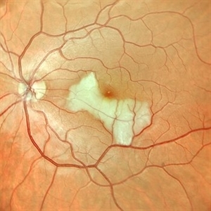
Color image capturing the left eye of a 32-year-old female. Despite a negative ophthalmological and medical history, she reported three days of blurred vision and a paracentral scotoma in her left eye, while maintaining central vision. The image reveals retinal whitening, extends from the parafoveal region to the inferotemporal arcade indicative of cilioretinal artery occlusion. Following this observation, the patient was referred for systemic assessment to explore the underlying etiology of the occlusion.
Photographer: Eloy Mata-Cortes, MD, Instituto Mexicano de Oftalmología, Querétaro, México
Imaging device: Nidek Mirante
Condition/keywords: cilioretinal artery occlusion, oclussion, retinal whitening
-
 Retinal Ischemia, Edema, and Hemorrhages on the Infero-Temporal Macula
Aug 26 2019 by Narciso F. Atienza, MD, MBA, FASRS, FPCS, FPAO.
47-year-old female who came in with blurring of vision of the right eye of 2 weeks duration. She is hypertensive with poor control, taking Amlodipine irregularly. Denies any cardiac problem non-diabetic. Vision upon presentation was 20/400 (OD), 20/20 (OS) colored fundus photo of the right eye showing areas of retinal ischemia, edema and hemorrhages on the infero-temporal macula extending to the arcade.
Photographer: Narciso F Atienza, Jr. MD, MBA
Imaging device: Topcon TRC
Condition/keywords: edema, hemorrhage, inferotemporal arcade, retinal ischemia
-
 CMV Retinitis/ Before Treatment
Mar 13 2015 by Niloofar Piri, MD
Fundus photograph of the left eye of a 40-year-old Caucasian female with history of positive HIV test for 23 years. She has been off HAART therapy for the past 2 years and presented with decreased vision OS and upper visual field defect. On examination, she had trace cells in anterior vitreous , hemorrhagic retinitis which starts around the optic nerve and extending to inferotemporal arcade with secondary inferotemporal BRVO; in temporal periphery , she had granular pattern of CMV retinitis which is a manifestation of outer retina involvement.
Photographer: Angela Anderson
Condition/keywords: CMV retinitis, HIV
 Loading…