Search results (14 results)
-
 A Large Break at the Posterior Pole With RD With PVR (S/p Old Blunt Trauma)
Jan 16 2025 by Anand Temkar
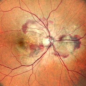
Right eye widefield fundus color photo of a 10 year old kid who noticed diminution of vision in right eye since a month. We can see the large break at the posterior pole with rolled up margins associated with retinal detachment and PVR changes.
Photographer: Dr.Anand Temkar- Retina Foundation, Ahmedabad
Imaging device: Mirante
Condition/keywords: posterior pole break, proliferative vitreoretinopathy (PVR), Retinal Detachment
-
 Right Eye Color Photo With Hemorrhages in Case of CNVM With Angioid Streaks
Nov 29 2024 by Anand Temkar
A 45 year old male came with chief complaint of blurring vision in right eyes since past 4 days. His vision is 6/12 in right eye and 6/9 in left eye. His vision was 14 mmHg in right eye and 16 mmHg in left eye. He was diagnosed with Angioid Streaks in both eyes about a year ago, then he developed choroidal neovascularization in his left eye 8 months ago, for which he received AntiVEGF injections x 3. Left eye is a stable eye now. Patient presented with right eye choroidal neovascularization in a case of Angioid Streaks on recent follow up. We have advised him right eye AntiVEGF injections x 3. In this image, the right eye color photo shows bleed from CNVM in case of angioid streaks.
Photographer: Dr.Anand Temkar- Retina Foundation, Ahmedabad
Imaging device: Mirante
Condition/keywords: Angioid Streaks, choroidal neovascular membrane (CNVM)
-
 Dislocated Lens, Posterior OD
Jan 26 2024 by Corey Grant
OPTOS California photo presents a 71 year old male patient with a dislocated lens, posterior in the right eye. Presented on 1/26/24 with posteriorly dislocated SN60WF with a Soemmerring ring. Associated retinal hemorrhage within retinoschisis as well. This will result in a PPV/IOL exchange/SFIOL/STK for the right eye.
Photographer: Corey Grant, Ophthalmic Imager, Retina Specialist of Michigan
Imaging device: OPTOS California
Condition/keywords: color photo, IOL, OD, Optos, OPTOS CALIFORNIA, pars plana vitrectomy (PPV), retina
-
 Total retinal Detachment multiple holes
Sep 26 2022 by Denica Rodriguez
60 year old Male presented with two week old Macula off Retinal detachment with multiple tears.
Photographer: Denica Rodriguez
Imaging device: Optos California
Condition/keywords: color fundus photograph, color photo, macula-off, optos, pseudocolor, Retinal detachment, retinal holes, retinal tear, Retinal tear with detachment, superior arcade, superior field, superior retina, total retinal detachment
-
 Prepapillary Vascular Loop
Mar 11 2020 by Asdrubal F Moreno, MD
Fundus color photograph of a 80-year-old woman with a unilateral congenital prepapillary vascular loop and hypertensive retinopathy, focused on the retinal plane for perception.
Photographer: Asdrubal Moreno, Fundacion AVAO, Universidad de Los Andes, Venezuela
Imaging device: Zeiss Visucam 500
Condition/keywords: congenital prepapillary vascular loop, peripapillary
-
 Macular Pucker With Myelinated Nerve Fiber Layer
Nov 1 2018 by Kevin J. Blinder, MD, FASRS
Multi-color photo of macular pucker with myelinated nerve fiber layer.
Photographer: Jarrod Wehmeier
Imaging device: Heidelberg Spectralis
Condition/keywords: macular pucker
-
 Retinal Detachment
Oct 25 2018 by Graciela Nahuelquín Ríos
A 45-year-old patient reported a blow to the right eye 1 month ago, and a week ago he presented with low visual acuity. In retinal mapping and background color photography retinal detachment with giant rupture in temporal arch.
Photographer: Lic. TM. Graciela Nahuelquín Ríos
Imaging device: TRC-50DX - Topcon
-
 Retinal Detachment
Feb 17 2018 by JEFFERSON R SOUSA, Tecg.º (Biomedical Systems Technology)
A 42-year-old patient complained of low vision in the left eye. In retinal mapping and background color photography, extensive retinal detachment was observed.
Photographer: JEFFERSON R SOUSA - Study Center and Ophthalmological Research Dr. Andre M V Gomes, Institute Dr. Suel Abujamra São Paulo-Brazil
Imaging device: Fundus camera Topcon TRC-50 DX, Imaginet 5.0, angle de 50 graus. Flash 36 / Mosaic with 10 images.
-
 Autosomal Recessive Bestrophinopathy - Color Photo OD
Dec 22 2017 by Tony Tsai, MD, FASRS
11-year-old Asian male with 20/40 vision OU, negative family history for ocular conditions, and bilateral atypical vitelliform deposits and subretinal fluid. EOG confirmed abnormally low Arden ratios OU. Genetic testing revealed homozygous recessive mutation in BEST1 gene (p.L140V:c.418C>G). Also known as p.L80V; Ref: Davidson (2009) Am J Hum Genet 85, 581.
Photographer: San Juanita Zazueta
Imaging device: Topcon
Condition/keywords: Best disease
-
 Chronic Inferior Retinal Detachment
Mar 1 2017 by Philip J. Polkinghorne, MD
Color photograph of chronic retinal detachment with pigment demarcation line and atrophic holes visible. The vision was recorded at 20/20, and follow up is 3 years.
Photographer: Alex Fraser
Condition/keywords: atrophic retinal hole, demarcation line
-
 Degeneration Paravenous
Sep 20 2016 by JEFFERSON R SOUSA, Tecg.º (Biomedical Systems Technology)
Female patient, 32-years-old, Asian, appeared at the clinic with a history of glaucoma. 20/20 visual acuity in both eyes. Examination of color photography, pigmentary changes were observed following the vascular arcades only in the left eye. Suggestive of paravenous degeneration.
Photographer: JEFFERSON R SOUSA - Study Center and Ophthalmological Research Dr. Andre M V Gomes, Institute Dr. Suel Abujamra São Paulo-Brazil
Imaging device: Zeiss / VisuCam-500 - Angulation of field photo of 45 Degrees, flash 24.
Condition/keywords: degeneration paravenous
-
 Retinoschisis Right Eye
Oct 27 2014 by AnneMarie Smykowski
65-year-old white male, with hypertensive retinopathy. He has a stable retinoschisis for approximately 10 years.
Photographer: AnneMarie Smykowski C.O.A., Island Retina Shirley, NY
Imaging device: Optos Daytona
Condition/keywords: color photo, Daytona, Optos, retinoschisis
-
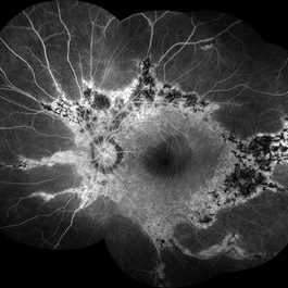
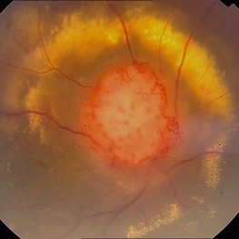 Color Photo of Optic Disc Capillary Hemangioblastoma
Mar 18 2014 by Arwa Azmeh, MD, PhD
Color fundus photograph of an 48-year-old male who complained of decreased visual acuity in his right eye over the last few months. Systemically the patient was healthy. His VA was OD Cf 3m, OS 20/20. Anterior segments were WNL in OU. IOP was WNL in OU. Fundus exam OD revealed unpigmented mass over the optic disc with retinal venous tortuosity at its edges with a ring of thick HYE surrounding it and shallow RD in this area extending to the foveal area. Several few small retinal hemorrhages were seen in the far retinal periphery which were explained to be caused by venous stasis due the optic disc tumor.
Condition/keywords: color photo, optic disc, retinal hemangioblastoma
-
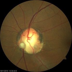 Optic Nerve Coloboma With 2 Pits, Nasal and Temporal Color
Nov 21 2013 by Alexandre Durao Alves Pereira, MD
Fundus photograph, color, red free, blue lite and FAF of a optic nerve coloboma with 2 pits, one nasal and other temporal.
Photographer: Alexandre Pereira
Imaging device: Visucam 300
Condition/keywords: color photo, optic nerve coloboma
 Loading…