Search results (8 results)
-
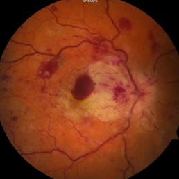
 Combined central retinal vein occlusion and branch retinal arteriolar occlusion
Sep 13 2022 by Ruchir Mehta, DO, DNB, FRCS
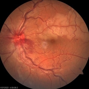
Fundus photograph of left eye of a 63 years old female with known type 2 DM and HTN showing combined central retinal venous occlusion and superior branch retinal arteriolar occlusion
Photographer: Ruchir Mehta, Mehta Superspeciality Eye Hospital, Jamnagar, Gujarat, India
Imaging device: Zeiss Visucam 500
Condition/keywords: branch retinal artery occlusion (BRAO), central retinal vein occlusion (CRVO), COMBINED
-
 Venous Beading
Nov 4 2021 by Stefanie Palmer
Venous Beading in a patient with both PDR and CRVO.
Photographer: Stefanie Palmer, CRA
Imaging device: Topcon
Condition/keywords: central retinal vein occlusion (CRVO), diabetic retinopathy, proliferative diabetic retinopathy (PDR), venous beading
-
 Central Retinal Vein Occlusion with Cilioretinal Artery Occlusion
Oct 21 2020 by Rutul R Patel, MD Ophthalmology
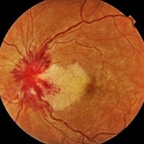
Fundus photograph of left eye of 37-year-old female who presented with sudden painless loss of vision in left eye due to CRVOwith CLRAO.
Photographer: Vidhi Bavishi, Shivjyoti Eye Hospital
Imaging device: TOPCON MAESTRO
Condition/keywords: central retinal vein occlusion (CRVO), cilioretinal artery occlusion
-
 CRVO with Secondary CLRAO
May 28 2020 by Richard M Martindale, MD
Non-ischemic CRVO (VA 20/30) with secondary CLRAO (nasal macular pallor) in a hypertensive 69yo female. Pathophysiologically, the cilioretinal artery occlusion occurs secondary to elevation in the hydrostatic pressure in the retinal venous system relative to the choroidal perfusion pressure (which supplies the cilioretinal artery).
Photographer: Retina Consultants of Alabama, P. C.
Imaging device: Optos
Condition/keywords: cilioretinal artery occlusion, non-ischemic central retinal vein occlusion (CRVO)
-
 Ischemic Central Retinal Vein Occlusion
Jan 24 2019 by Nichole Lewis
76-year-old woman with an ischemic central retinal vein occlusion, severely attenuated and sclerotic vessels and scattered retinal hemorrhages. Vision decrease over 1 year. VA 20/CF. Patient is returning for pan retinal photocoagulation.
Photographer: Nichole Lewis
Imaging device: Optos
Condition/keywords: attenuated vessels, central retinal vein occlusion (CRVO), hemorrhage, ischemic CRVO, sclerotic vessels
-
 CRVO
CRVO
Apr 22 2017 by Gabriel Costa Andrade, PhD
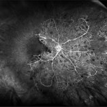
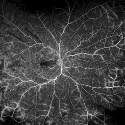
Panoramic retinography (Optos® California) of the right eye of a 48-year-old female patient with a history of low-vision in the right eye 2 months ago. At the exam presented visual acuity of 20/200 in the right eye and 20/20 in the left eye. Angiography shows diffuse perivascular leakage associated with areas of hypoperfusion in macula and periphery.
Photographer: Gabriel Andrade
Imaging device: Optos® California
Condition/keywords: central retinal vein occlusion (CRVO)
-
 Branch Retinal Artery Occlusion With Concurrent Central Retinal Vein Occlusion
Oct 5 2016 by Larry M Puthenparambil, MD
Branch retinal artery occlusion with concurrent central retinal vein occlusion.
Photographer: Stacey Groom
Imaging device: Topcon
Condition/keywords: branch retinal artery occlusion (BRAO), central retinal vein occlusion (CRVO)
-
 Non-Ischemic Central Retinal Vein Occlusion
Jun 28 2016 by Nichole Lewis
53-year-old female with non-ischemic central retinal vein occlusion left eye.
Photographer: Nichole Lewis
Condition/keywords: non-ischemic central retinal vein occlusion (CRVO)
 Loading…