Search results (10 results)
-
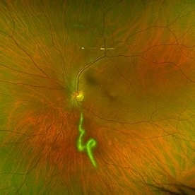 Vitreous Cavity Inhabitant
Jun 2 2025 by Poornachandra B, MS, FVRS
A 36-year-old male presented with a 6-week history of intermittent ocular redness, now accompanied by the recent onset of floaters for the past 2 days. Fundus examination revealed the presence of a nematode in the vitreous cavity.
Photographer: Mr Dhikshith
Condition/keywords: parasite
-
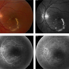
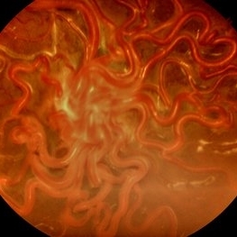 Wyburn-Mason Syndrome (Racemose Angioma)
Mar 23 2024 by Pushkar Mahale
Fundus photograph of a 10 year old child presenting with no perception of light in right eye. Fundus examination revealed dilated and tortuous retinal vessels suggestive of Racemose Hemangioma.
Photographer: Dr Pushkar Mahale
Condition/keywords: racemose hemangioma, Wyburn -Mason Syndrome
-
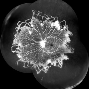 Vascular Non Perfusion in Takayasu Arteritis
Feb 6 2024 by SHILPI H NARNAWARE, ICO ( Retina) , FAICO ( Vitreo-Retina)
A case of 16 year-old female with combined RD in RE. Fundus examination & FFA revealed 360 degrees non-perfusion in periphery in non-symptomatic eye.
Photographer: Shilpi Narnaware, Sarakshi Netralaya , Nagpur, Maharashtra , India
Imaging device: Mirante ( by Nidek)
Condition/keywords: CNP areas, takayasu arteritis
-
 Ocular Hypotony Due to Leaking Bleb
Apr 1 2019 by Anfisa Ayalon, MD
81-year-old male who had trabeculectomy in his right eye 4 years ago, presented to the emergency room with complains of decreased vision in that eye for two months. Slit-lamp examination showed cystic bleb with leakage, intraocular pressure was 0 MMHg. Fundus examination showed hypotony maculopathy, peripheral choroidal detachments, multiple chorioretinal folds with subretinal fluid.
Photographer: Anfisa Ayalon, MD., Meir Medical Center, Kfar Saba, Israel.
Imaging device: California, Optos 200 DTX
Condition/keywords: choroidal detachment, hypotonous retinopathy, hypotony maculopathy
-
 Combined Hamartoma
Feb 29 2016 by Andrea Arriola-Lopez, MD MSc
40 year-old man with diminished VA since 6 month ago. Fundus examination revealed macular folds, yellow-whitish elevated lesion at the fovea and a subretinal hemorrhage.
Photographer: Andrea Elizabeth Arriola-Lopez MD, MSc
Imaging device: OPTOS Dakota
Condition/keywords: combined hamartoma, macula, subretinal hemorrhage
-
 Vascular Anormalities
Jan 6 2016 by Andrea Arriola-Lopez, MD MSc
77-year-old man. Decrease of visual acuity OS. VA 20/30 IOP 14mmHg. Fundus examination findings: Hard exudates, microaneurysms near to fovea. OCT shows IRF. Late leakage on FA.
Photographer: Andrea Elizabeth Arriola-Lopez, MSc MD
Condition/keywords: abnormal retinal vessel, aneurysm, hard exudates, vascular anomaly
-
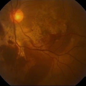 Chorioretinitis Sclopetaria
Jan 22 2016 by Jorge Morales-Martínez, MD
Fundus photograph of a 27-year-old male that sustained a traumatic injury in his left eye with a paintball projectile. Fundus examination showed a large subretinal hemorrhage, areas of commotio retinae and maculopathy.
Photographer: Jorge Morales-Martínez MD
-
 APMPPE in a 21 Year-Old Female Patient
Oct 23 2015 by Roy Schwartz, MD
FA photograph of a 21-year-old, usually healthy, female, presenting with visual deterioration and photophobia in BE. Upon examination deep lesions were seen on fundus examination.
Photographer: Galit Yair Pur
Condition/keywords: acute posterior multifocal placoid pigment epitheliopathy (APMPPE)
-
 Chronical Submacular Hemorrhage in the Setting of Neovascular AMD
Mar 23 2015 by Rita Couceiro, MD, MS
An 80-year-old male, with a history of hypertension and high cholesterol, complained of acute and painless vision loss in his left eye (OS) in the previous 5 months. On observation best corrected visual acuity in OS was hand motion. A dense vitreous opacity in OS precluded fundus examination. Ocular ultrasound revealed vitreous hemorrhage and thickening of the macular area. The patient was submitted to pars plana vitrectomy, which disclosed a large submacular hemorrhage with chronical features and disciform scarring in the setting of neovascular AMD.
Imaging device: Intraoperative fundus photograph
Condition/keywords: neovascular age-related macular degeneration (AMD), submacular hemorrhage, wet age-related macular degeneration (wet AMD)
-
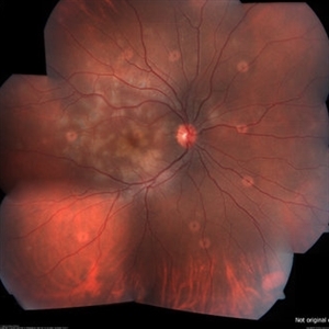
000---thumb.jpg/image-square;max$300,300.ImageHandler) Fundus Panorama Finding of Tractional Retinal Detachment Due to Proliferative Diabetic Retinopathy
Fundus Panorama Finding of Tractional Retinal Detachment Due to Proliferative Diabetic Retinopathy
Dec 25 2013 by Dong Yoon Kim, MD
47-year-old woman visited our clinic for decreased visual acuity on her right eye. Her visual acuity of right eye was hand motion. Fundus examination showed traction retinal detachment.
Condition/keywords: fundus photograph, tractional retinal detachment
 Loading…