Search results (6 results)
-
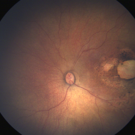
 Macular Colobomas in Congenital Zika Syndrome
Sep 26 2020 by Swati Agarwal-Sinha, MD, FASRS
Color fundus picture of the right (OD) and left (OS) eye of 3-day-old female infant with congenital Zika syndrome with bilateral macular colobomatous like chorioretinal atrophy, attenuated vessels, pigmentary changes, and optic disc pallor.
Photographer: Swati Agarwal-Sinha, MD
Condition/keywords: fundus photograph, macular coloboma, zika
-
 ZIKA
ZIKA
May 8 2018 by Audina M. Berrocal, MD FASRS
ZIKA
Photographer: Brenda Fallas
Imaging device: RetCam
Condition/keywords: zika
-
 ZIKA
ZIKA
May 8 2018 by Audina M. Berrocal, MD FASRS
ZIKA
Photographer: Brenda Fallas
Imaging device: retCam
Condition/keywords: zika
-
 Zika
Zika
Oct 13 2017 by Carmen R. Negrin-Martin, MD
5-month-old baby boy; Zika virus.
Photographer: Rosina Negrín, MD, Santo Domingo , Dominican Republic
Imaging device: Inview volk
Condition/keywords: zika
-
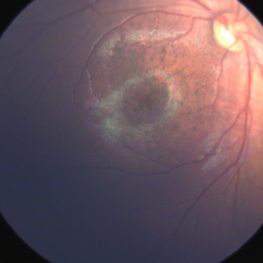
 Congenital Zika Syndrome
Jun 29 2017 by Camila V Ventura, MD, PhD
Infant with congenital zika syndrome presenting with: two macular chorioretinal scars, and pigment mottling in the macula and inferior temporal arcade.
Photographer: Camila Ventura, MD - Altino Ventura Foundation, Brazil
Imaging device: RetCam®
Condition/keywords: chorioretinal atrophy, chorioretinal scar, focal pigmentary changes, pigment mottling
-
 Congenital Zika Syndrome
Jun 29 2017 by Camila V Ventura, MD, PhD
Fundus image of an infant with congenital zika syndrome presenting with focal pigmentary changes in the macular region
Photographer: Camila Ventura, MD - Altino Ventura Foundation, Brazil
Imaging device: Retcam®
Condition/keywords: focal pigmentary changes
 Loading…