Search results (45 results)
-
---thumb.jpg/image-square;max$300,300.ImageHandler) Age Related Macular Degeneration
Age Related Macular Degeneration
May 3 2013 by Suber S. Huang, MD, MBA, FASRS
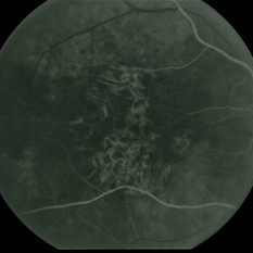
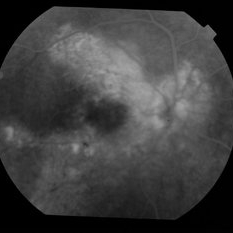
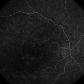
Age related macular degeneration.
Condition/keywords: advanced geographic atrophy, atrophic scar, atrophic spot, geographic atrophy, macula lesion, pigment epithelial atrophy, red-free, window defect
-
---thumb.jpg/image-square;max$300,300.ImageHandler) Age Related Macular Degeneration - Geographic Atrophy
Age Related Macular Degeneration - Geographic Atrophy
May 3 2013 by Suber S. Huang, MD, MBA, FASRS
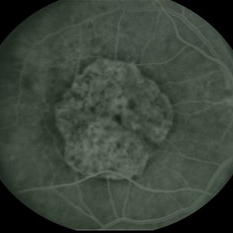
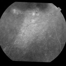
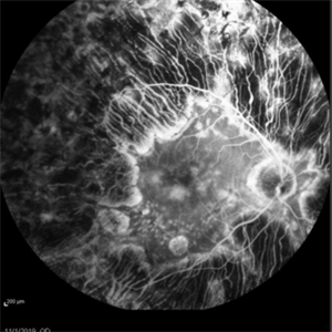
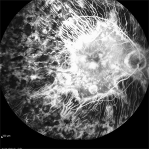
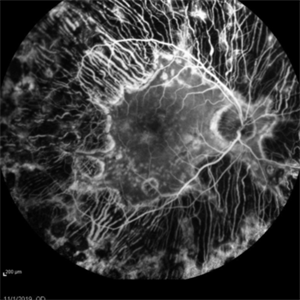
Geographic Atrophy.
Imaging device: Retina Diseases Imaging Reading Center
Condition/keywords: advanced geographic atrophy, atrophic scar, atrophic spot, geographic atrophy, macula lesion, pigment epithelial atrophy, red-free, window defect
-
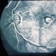 FA After Thermal Laser for CNVM
Feb 19 2015 by H. Michael Lambert, MD
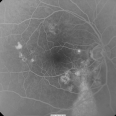
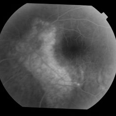
AV phase of fluorescein angiogram with RPE window defect after thermal laser.
Condition/keywords: choroidal neovascular membrane (CNVM), laser, window defect
-
 Idiopathic Uveal Effusion Syndrome
Aug 22 2024 by Jordyn Beckman
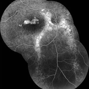
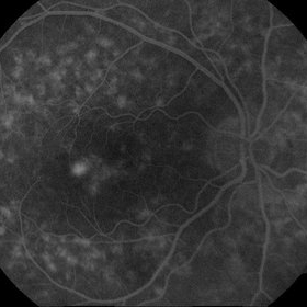
61 year old male with Idiopathic Uveal Effusion Syndrome with starry night appearance on fluorescein. 3 weeks s/p single external drainage retinotomy and 9 weeks of oral pred with recurrent choroidal effusions. Has since returned to surgery for secondary drainage retinotomy; subretinal fluid remain persistent.
Photographer: Jordyn Beckman
Imaging device: Optos California
Condition/keywords: chorioretinitis, Choroidal, exudative detachment, window defect
-
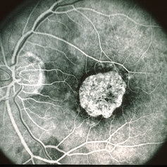
 Adenocarcinoma Arising from CHRPE
Sep 17 2015 by Marc C. Peden, MD
49-year-old female referred for presumed ocular melanoma. On examination was noted to have darkly pigmented lesion in the temporal retina of left eye. Lesion had characteristic scalloped edges with central lacunae, however, on ultrasonography was noted to have 1.8mm of elevation with high internal reflectivity. IVFA shows absence of dual circulation with areas of window defect. Findings were consistent with those described by Shields et al., in their April 2001 article in Archives of Ophthalmology.
Photographer: Janet Traynom
Imaging device: Optos P200MA
Condition/keywords: adenocarcinoma arising from CHRPE
-
 Adenocarcinoma Arising from CHRPE
Sep 17 2015 by Marc C. Peden, MD
49-year-old female referred for presumed ocular melanoma. On examination was noted to have darkly pigmented lesion in the temporal retina of left eye. Lesion had characteristic scalloped edges with central lacunae, however, on ultrasonography was noted to have 1.8mm of elevation with high internal reflectivity. IVFA shows absence of dual circulation with areas of window defect. Findings were consistent with those described by Shields et al., in their April 2001 article in Archives of Ophthalmology.
Photographer: Janet Traynom COT
Imaging device: Optos P200MA
Condition/keywords: adenocarcinoma arising from CHRPE
-
 Alports Disease
Jul 29 2013 by H. Michael Lambert, MD
Alports disease, macular diseases not associated with FA changes. Spotty window defects with mid peripheral lesions. Disease may represent ABNL in basement membranes. Basal laminar drusen.
Condition/keywords: Alports disease
-
 Alports Disease
Jul 29 2013 by H. Michael Lambert, MD
Alports disease, macular diseases not associated with FA changes. Spotty window defects with mid peripheral lesions. Disease may represent ABNL in basement membranes. Basal laminar drusen.
Condition/keywords: Alports disease
-
 ARMD with RPE Rip
Oct 12 2012 by Jeffrey G. Gross, MD, FASRS
ARMD with RPE rip, FA, showing window defect and blockage from retracted RPE layer.
Condition/keywords: retinal pigment epithelium, retinal pigment epithelium (RPE) tear, retracted retinal pigment epithelium (RPE) layer
-
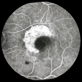
 Bull's Eye
May 2 2013 by Henry J. Kaplan, MD
Fluorescein angiography demonstrates RPE window defects and hyperfluorescence in bull's eye maculopathy; #2.
Condition/keywords: bull's eye maculopathy
-
 Central areolar atrophy
May 2 2013 by Henry J. Kaplan, MD
Fluorescein angiogram demonstrates early hyperfluorescence due to window defect; the patient is young with GA like lesion and vision of 20/200 which is compatible with central areolar atrophy#1
Condition/keywords: central areolar choroidal dystrophy (CACD)
-
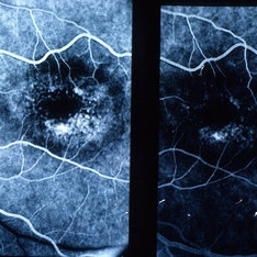
 Central Areolar Choroidal Dystrophy
Jan 5 2015 by H. Michael Lambert, MD
Early fluorescein angiogram OD with central, well defined, confluent atrophy, window defect with choroidal vessels visible.
Condition/keywords: central areolar choroidal dystrophy (CACD)
-
 Central Areolar Choroidal Dystrophy
Jan 5 2015 by H. Michael Lambert, MD
Later fluorescein angiogram OD with central, well defined, confluent atrophy, window defect with transmission.
Condition/keywords: central areolar choroidal dystrophy (CACD)
-
 Chronic Central Serous Chorioretinopathy
Oct 31 2012 by Lihteh Wu, MD
FA frame showing a hyperfluorescent window defect in a gutter pattern. There is also a hot spot in the nasal macula.
-
 Chronic Central Serous Chorioretinopathy
Oct 31 2012 by Lihteh Wu, MD
FA frame showing a hyperfluorescent window defect in a gutter pattern extending down from the posterior.
Condition/keywords: central serous chorioretinopathy (CSCR)
-
 Chronic CSCR - RPE Tracts
May 4 2014 by Neha Goel, MS DNB FRCS (Glasg)
FFA of a patient with chronic CSCR showing RPE window defects at the macula and RPE tracts running inferiorly from the peripapillary region.
Photographer: Neha Goel
Imaging device: Zeiss Visucam
Condition/keywords: chronic central serous chorioretinopathy (CSCR), retinal pigment epithelium
-
 Chronic CSCR Resolution With Anti-VEGF
Jul 31 2014 by Mallika Goyal, MD
Fluorescein of the right eye of a 55-year-old male who presented with symptoms from chronic CSCR (> 3 years) shows extensive RPE window defects and occasional areas of intense hyperfluorescence.
Photographer: Mallika Goyal, MD, Apollo Health City, Jubilee Hills, Hyderabad-500033
Condition/keywords: chronic central serous chorioretinopathy (CSCR)
-
 Chronic CSCR Resolution With Anti-VEGF
Jul 31 2014 by Mallika Goyal, MD
Fluorescein of the inferior fundus of the right eye of a 55-year-old male who presented with symptoms from chronic CSCR (> 3 years) shows extensive RPE window defects and occasional areas of intense hyperfluorescence.
Photographer: Mallika Goyal, MD, Apollo Health City, Jubilee Hills, Hyderabad-500033
Condition/keywords: chronic central serous chorioretinopathy (CSCR)
-
 Chronic CSCR Resolution With Anti-VEGF
Jul 31 2014 by Mallika Goyal, MD
Fluorescein of the left eye of a 55-year-old male who presented with fellow eye symptoms from chronic CSCR (> 3 years) shows extensive RPE window defects.
Photographer: Mallika Goyal, MD, Apollo Health City, Jubilee Hills, Hyderabad-500033
Condition/keywords: chronic central serous chorioretinopathy (CSCR)
-
 CNV due to AMPPE
Oct 16 2012 by Ratimir Lazic, MD, PhD
FAG of 58-year-old male. In late venous phase hyperflorescence of white dots (caused by window defect) can be seen. Intensive leakage of dye in juxtafoveolar region.
Photographer: Marko Lukic, MD
Imaging device: Zeis Visucam Lite 2
Condition/keywords: acute posterior multifocal placoid pigment epitheliopathy (APMPPE), choroidal neovascularization (CNV)
-
 CNV due to AMPPE
Oct 16 2012 by Ratimir Lazic, MD, PhD
FAG of 58-year-old male. In early venous phase hyperflorescence of white dots (caused by window defect) can be seen. Leakage of dye in juxtafoveolar region.
Photographer: Marko Lukic, MD
Imaging device: Zeis Visucam Lite 2
Condition/keywords: acute posterior multifocal placoid pigment epitheliopathy (APMPPE), choroidal neovascularization (CNV)
-
---thumb.jpg/image-square;max$300,300.ImageHandler) Cone Dystrophy
Cone Dystrophy
Feb 20 2013 by From the Collections of Thomas M. Aaberg, MD and Thomas M. Aaberg Jr., MD
FA of OS showing window defects in a circular pattern at the macula.
Condition/keywords: bull's eye maculopathy, cone dystrophy
-
 Enough PRP? (Proliferative Diabetic Retinopathy With Extreme PRP and Widespread Atrophy)
Nov 24 2019 by Thomas A. Ciulla, MD, MBA, FASRS
Fluorescein angiogram from a 71-year-old woman who underwent numerous sessions of pan retinal laser photocoagulation for proliferative diabetic retinopathy in the remote past. Note the widespread severe secondary atrophy, with only the central macular RPE remaining. Note the choroidal vessels through the diffuse window defect in the peripheral macula and near periphery.
Condition/keywords: laser injury, laser photocoagulation, proliferative diabetic retinopathy (PDR)
 Loading…