Search results (20 results)
-
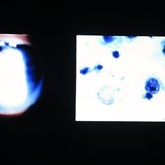 Slide 8-27
Mar 4 2019 by Lancaster Course in Ophthalmology
Ocular reticulum-cell sarcoma. Appearance of vitreous cells and debris (left) in a 61-year-old man with a 2-year history of recalcitrant uveitis. Pars plana vitrectomy specimen was prepared, using a millipore filter and staining by a modified Papanicolaou technique. Tumor cells with nuclear cytoplasmic disproportion and finger like nuclear extensions are characteristic of reticulum-cell sarcoma (right). (E.P. No. 38606)
Condition/keywords: pars plana vitrectomy (PPV), reticulum cell sarcoma, uveitis, vitreous cells
-
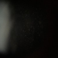 Intermediate Uveitis
Dec 14 2018 by Hashim Ali Khan, OD, FAAO
Vitreous cells in a 30-year-old female with intermediate uveitis.
Condition/keywords: vitritis
-
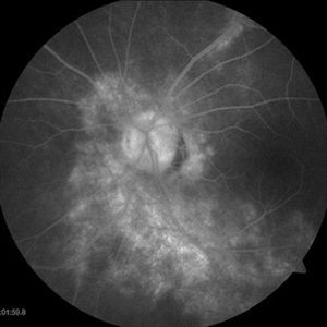
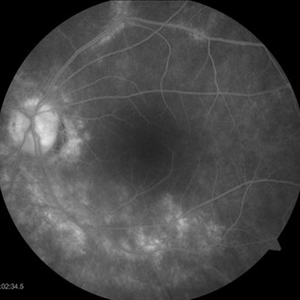
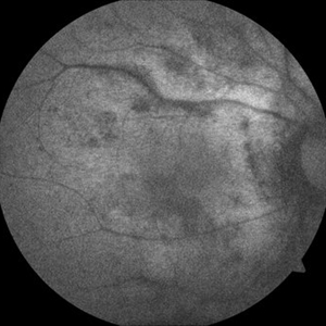
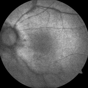
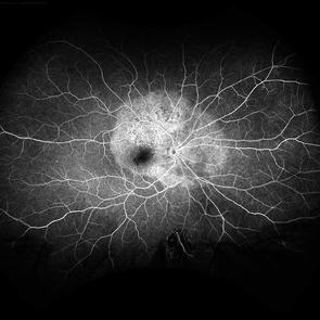 Acute Syphilitic Posterior Placoid Chorioretinitis
May 4 2021 by RAFAEL REIS PEREIRA, MD
A 31-year-old patient with a complaint of photophobia and low visual acuity OD in the previous three weeks. BCVA was 20/60 and 20/20 The fundus examination revealed a placoid white lesion in the posterior pole and vitreous cells in the right eye. The left eye was unremarkable. Fluorescein angiography reveals hyperfluorescent plaque with distinctive “leopard spots” hypofluorescence.
Imaging device: Opto California
Condition/keywords: acute syphilitic posterior placoid chorioretinitis
-
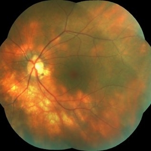
---thumb.jpg/image-square;max$300,300.ImageHandler) Birdshot Case #1 OD Color
Birdshot Case #1 OD Color
May 1 2013 by Armando L. Oliver, MD
64-year-old Puerto Rican woman consulted due to the presence of 1+ vitreous cells. The fundus examination revealed orange to yellow lesions dispersing from the disk. Work-up revealed she was HLA-A29 positive and the suspected diagnosis of Birdshot Chorioretinopathy was made. Chest X-Ray, FTA-Abs and RPR were negative.
Photographer: Moises Castro, Instituto de Ojos y Piel, Carolina, PR
Imaging device: Zeiss, Visucam NM/FA
Condition/keywords: birdshot, birdshot chorioretinopathy, birdshot retinochoroidopathy
-
---thumb.jpg/image-square;max$300,300.ImageHandler) Birdshot Case #1 OD FAF
Birdshot Case #1 OD FAF
May 1 2013 by Armando L. Oliver, MD
64-year-old Puerto Rican woman consulted due to the presence of 1+ vitreous cells. The fundus examination revealed orange to yellow lesions dispersing from the disk. Work-up revealed she was HLA-A29 positive and the suspected diagnosis of Birdshot Chorioretinopathy was made. Chest X-Ray, FTA-Abs and RPR were negative.
Photographer: Moises Castro, Instituto de Ojos y Piel, Carolina, PR
Imaging device: Zeiss, Visucam NM/FA
Condition/keywords: birdshot, birdshot chorioretinopathy, birdshot retinochoroidopathy
-
---thumb.jpg/image-square;max$300,300.ImageHandler) Birdshot Case #1 OD IVFA
Birdshot Case #1 OD IVFA
May 1 2013 by Armando L. Oliver, MD
64-year-old Puerto Rican woman consulted due to the presence of 1+ vitreous cells. The fundus examination revealed orange to yellow lesions dispersing from the disk. Work-up revealed she was HLA-A29 positive and the suspected diagnosis of Birdshot Chorioretinopathy was made. Chest X-Ray, FTA-Abs and RPR were negative.
Photographer: Moises Castro, Instituto de Ojos y Piel, Carolina, PR
Imaging device: Zeiss, Visucam NM/FA
Condition/keywords: birdshot, birdshot chorioretinopathy, birdshot retinochoroidopathy
-
 Birdshot Case #1 OS Color
May 1 2013 by Armando L. Oliver, MD
64-year-old Puerto Rican woman consulted due to the presence of 1+ vitreous cells. The fundus examination revealed orange to yellow lesions dispersing from the disk. Work-up revealed she was HLA-A29 positive and the suspected diagnosis of Birdshot Chorioretinopathy was made. Chest X-Ray, FTA-Abs and RPR were negative.
Photographer: Moises Castro, Instituto de Ojos y Piel, Carolina, PR
Imaging device: Zeiss, Visucam NM/FA
Condition/keywords: birdshot, birdshot chorioretinopathy, birdshot retinochoroidopathy
-
---thumb.jpg/image-square;max$300,300.ImageHandler) Birdshot Case #1 OS Color
Birdshot Case #1 OS Color
May 1 2013 by Armando L. Oliver, MD
64-year-old Puerto Rican woman consulted due to the presence of 1+ vitreous cells. The fundus examination revealed orange to yellow lesions dispersing from the disk. Work-up revealed she was HLA-A29 positive and the suspected diagnosis of Birdshot Chorioretinopathy was made. Chest X-Ray, FTA-Abs and RPR were negative.
Photographer: Moises Castro, Instituto de Ojos y Piel, Carolina, PR
Imaging device: Zeiss, Visucam NM/FA
Condition/keywords: birdshot, birdshot chorioretinopathy, birdshot retinochoroidopathy
-
---thumb.jpg/image-square;max$300,300.ImageHandler) Birdshot Case #1 OS FAF
Birdshot Case #1 OS FAF
May 1 2013 by Armando L. Oliver, MD
64-year-old Puerto Rican woman consulted due to the presence of 1+ vitreous cells. The fundus examination revealed orange to yellow lesions dispersing from the disk. Work-up revealed she was HLA-A29 positive and the suspected diagnosis of Birdshot Chorioretinopathy was made. Chest X-Ray, FTA-Abs and RPR were negative.
Photographer: Moises Castro, Instituto de Ojos y Piel, Carolina, PR
Imaging device: Zeiss Visucam NM/FA
Condition/keywords: birdshot, birdshot chorioretinopathy, birdshot retinochoroidopathy
-
---thumb.jpg/image-square;max$300,300.ImageHandler) Birdshot Case #1 OS IVFA
Birdshot Case #1 OS IVFA
May 1 2013 by Armando L. Oliver, MD
64-year-old Puerto Rican woman consulted due to the presence of 1+ vitreous cells. The fundus examination revealed orange to yellow lesions dispersing from the disk. Work-up revealed she was HLA-A29 positive and the suspected diagnosis of Birdshot Chorioretinopathy was made. Chest X-Ray, FTA-Abs and RPR were negative.
Photographer: Moises Castro, Instituto de Ojos y Piel, Carolina, PR
Imaging device: Zeiss, Visucam NM/FA
Condition/keywords: birdshot, birdshot retinochoroidopathy
-
 Birdshot Case #1 OS IVFA
May 1 2013 by Armando L. Oliver, MD
64-year-old Puerto Rican woman consulted due to the presence of 1+ vitreous cells. The fundus examination revealed orange to yellow lesions dispersing from the disk. Work-up revealed she was HLA-A29 positive and the suspected diagnosis of Birdshot Chorioretinopathy was made. Chest X-Ray, FTA-Abs and RPR were negative.
Photographer: Moises Castro, Instituto de Ojos y Piel, Carolina, PR
Imaging device: Zeiss, Visucam NM/FA
Condition/keywords: birdshot, birdshot chorioretinopathy, birdshot retinochoroidopathy
-
 Birdshot Case #1 OS IVFA
May 1 2013 by Armando L. Oliver, MD
64-year-old Puerto Rican woman consulted due to the presence of 1+ vitreous cells. The fundus examination revealed orange to yellow lessions dispersing from the disk. Work-up revealed she was HLA-A29 positive and the suspected diagnosis of Birdshot Chorioretinopathy was made. Chest X-Ray, FTA-Abs and RPR were negative.
Photographer: Moises Castro, Instituto de Ojos y Piel, Carolina, PR
Imaging device: Zeiss, Visucam NM/FA
Condition/keywords: birdshot, birdshot chorioretinopathy, birdshot retinochoroidopathy
-
 Birdshot Case #1 OD FAF
May 1 2013 by Armando L. Oliver, MD
64-year-old Puerto Rican woman consulted due to the presence of 1+ vitreous cells. The fundus examination revealed orange to yellow lesions dispersing from the disk. Work-up revealed she was HLA-A29 positive and the suspected diagnosis of Birdshot Chorioretinopathy was made. Chest X-Ray, FTA-Abs and RPR were negative.
Photographer: Moises Castro, Instituto de Ojos y Piel, Carolina, PR
Imaging device: Zeiss, Visucam NM/FA
Condition/keywords: birdshot chorioretinopathy, birdshot retinochoroidopathy
-
 Birdshot Case #1 OS FAF
May 1 2013 by Armando L. Oliver, MD
64-year-old Puerto Rican woman consulted due to the presence of 1+ vitreous cells. The fundus examination revealed orange to yellow lesions dispersing from the disk. Work-up revealed she was HLA-A29 positive and the suspected diagnosis of Birdshot Chorioretinopathy was made. Chest X-Ray, FTA-Abs and RPR were negative.
Photographer: Moises Castro, Instituto de Ojos y Piel, Carolina, PR
Imaging device: Zeiss, Visucam NM/FA
Condition/keywords: birdshot, birdshot chorioretinopathy, birdshot retinochoroidopathy
-
 Cancer-Associated Retinopathy (CAR)
Jun 30 2018 by Peter G. Hovland, MD, PhD
Mosaic fundus photograph of affected right eye of 56-year-old woman 3 years after onset of cancer-associated retinopathy. Demonstrates RPE atrophy and attenuated retinal vasculature. Patient presented with vitreous cells.
Photographer: Colorado Retina Associates
Condition/keywords: retinopathy, vitreous
-
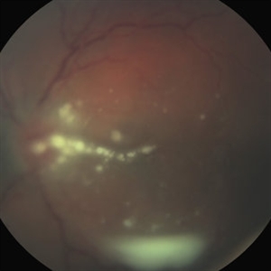 Fungal Endophthalmitis Associated With Intravenous Drug Abuse
Apr 16 2014 by Scott D. Schoenberger, MD
Fundus photograph of a 20-year-old male with pain and decreased vision OS for 3 days. His visual acuity was counting fingers and he had conjunctival injection, anterior chamber cells and vitreous cells. He admitted to intermittent use of intravenous heroin. A vitrectomy was performed and cultures were positive for candida albicans.
Condition/keywords: endogenous endophthalmitis, fungal endophthalmitis
-
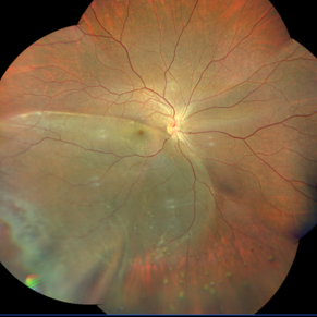 Inferior Temporal Dialysis-Retinal Detachment-RPE Vitreous Cells Clumps
Sep 2 2020 by Carlos W Arzabe, MD
Inferior temporal dialysis-retinal detachment-RPE vitreous cells clumps.
Imaging device: Clarus 700
Condition/keywords: retinal pigment epithelium
-
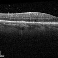 Ocular Syphilis
Feb 21 2024 by Nikhil K Bommakanti, MD
A monocular man in his sixties presented with blurred vision in the right eye for two months. Optical coherence tomography demonstrated vitreous cells and characteristic inflammatory deposits of the outer retina and retinal pigment epithelium, and laboratory testing confirmed the diagnosis of syphilis. He was admitted for intravenous penicillin and consultation with a specialist in infectious diseases.
Condition/keywords: syphilis
-
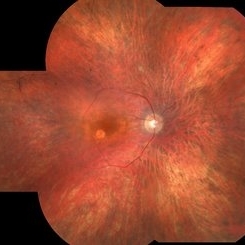
 Sympathetic Ophthalmia
Sep 28 2012 by Joseph M. Civantos, MD
59-year-old lady had blunt trauma left eye with a ruptured globe. She refused further surgery on that eye. She returned 4 months later with decreased vision in the right eye. RE dropped from 20/25 to 20/50. The view is hazy due to vitreous cells. Large white choroidal Dalen-Fuchs nodules are visible.
Condition/keywords: Dalen-Fuchs nodules, sympathetic ophthalmia
 Loading…