Search results (5 results)
-
 Bilateral Retinal Vaso-Occlusive Disease
Jun 20 2017 by S. Natarajan, MD, FASRS, FRCS (GLASGOW) , FICO, D.Sc, FELA
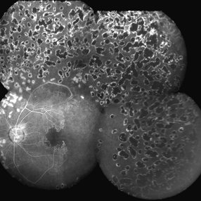
35-year-old woman with bilateral retinal vaso-occlusive disease with secondary neovascularization. She has undergone extensive care including photocoagulation to both the retina. There is severe ischemia on the posterior pole in both eyes. Patient has been started on immunosuppressant.
Photographer: Ms . Ashwini Borde
Imaging device: zeiss plus IR 450
Condition/keywords: neovascularization (NV), vaso-occlusive disease
-
 Eales Disease
Apr 1 2019 by Gary R. Cook, MD, FACS
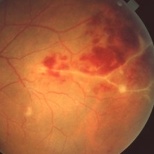
Fundus photograph of retinal vascular changes and retinal hemorrhages in the superotemporal periphery OS of a 23-year-old Vietnamese female with Eales disease; V.A. = 20/25-2.
Imaging device: Topcon VT-50
Condition/keywords: Eales disease, vaso-occlusive disease
-
 Eales Disease
Apr 1 2019 by Gary R. Cook, MD, FACS
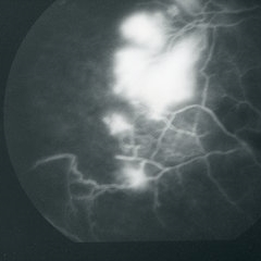
Late-phase fluorescein angiogram image of the retinal periphery of a 23-year-old Vietnamese female with Eales disease showing peripheral capillary nonperfusion, vaso-occlusion and peripheral retinal neovascularization; V.A.= 20/80.
Imaging device: Topcon VT-50
Condition/keywords: Eales disease, FA late phase, fluorescein angiogram (FA), peripheral retinal neovascularization, vaso-occlusive disease
-
 Eales Disease
Apr 1 2019 by Gary R. Cook, MD, FACS
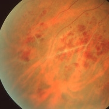
25-year-old Vietnamese male with peripheral retinal vasculitis, vaso-occlusion, and retinal hemorrhages secondary to Eales Disease; V.A.= 20/20.
Imaging device: Topcon VT-50
Condition/keywords: Eales disease, retinal hemorrhage, vaso-occlusive disease, vasoocclusive retinopathy
-
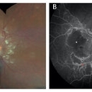 Retinal Lupus Vasculitis
Sep 25 2021 by Denis Jusufbegovic, MD
27-year-old woman with a history of systemic lupus erythematosus (SLE) presented with decreased vision to counting fingers at 2’ in the right eye. Funduscopic examination of the right eye (A) demonstrated retinal thickening and whitening of the macula, numerous cotton-wool spots, intra-retinal hemorrhages, sclerotic vessels, and vascular sheathing. Fluorescein angiography (B) demonstrated extensive vessel wall leakage (red asterisk) and large areas of capillary non-perfusion (white asterisk). These findings were consistent with severe vaso-occlusive retinopathy, a serious ophthalmologic manifestation of SLE. She was also diagnosed with concomitant cerebral lupus vasculitis. She was treated with intravenous methylprednisolone followed by oral prednisone taper and aspirin therapy. Mycophenolate mofetil was titrated to 1500 mg twice daily. Upon follow-up vision improved to 20/200.
Imaging device: Zeiss Clarus 500
Condition/keywords: cerebral lupus vasculitis, cotton wool spots, systemic lupus erythematosus (SLE) vasculitis, vaso-occlusive disease
 Loading…