Search results (16 results)
-
Ciliary Body Detachment in Uveal Effusion Syndrome
Apr 11 2025 by Siri Uppuluri
Ultrasound biomicroscopy of a phakic left eye in an 82-year-old man demonstrating ciliary body detachment in the setting of uveal effusion syndrome. Patient also presented with 360 choroidal effusions and underwent sclerectomy and drainage of choroidal effusions with resolution after surgical intervention.
Photographer: Siri Uppuluri, MD; Rutgers New Jersey Medical School
Condition/keywords: uveal effusion syndrome
-
 Uveal Effusion Syndrome
Mar 9 2013 by Gabriela Lopezcarasa Hernandez, MD
A 55-year-old man with glaucoma surgery and severe decrease in visual acuity, with superior nasal occlusion and serous retinal detachment.
Photographer: Araceli Rojas Arriaga, Hospital Angeles Lomas, Mexico
Imaging device: Zeiss FF4
Condition/keywords: idiopathic uveal effusion syndrome
-
 Uveal Effusion Syndrome
Oct 23 2023 by Gustavo Aguirre-Suarez
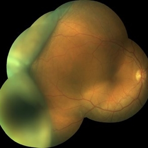
Fundus photograph of a 58-year-old man with Type 1 Uveal Effusion Syndrome, showing 360º bullous choroidal detachment.
Photographer: Dr. Gustavo Aguirre-Suarez
Imaging device: Zeiss Clarus 700
Condition/keywords: choroidal effusion, idiopathic uveal effusion syndrome
-
 Uveal Effusion Syndrome
Sep 19 2024 by Virginia Gebhart
61 year old female with idiopathic uveal effusion syndrome. 360 degrees of choroidal thickening, especially anterior with exudative fluid inferior. Mild vitritis present. Unable to gain venous access for FA, ultrasound and UBM performed which confirm choroidal and ciliary body thickening. Pt sent for inflammatory work up including MRI of brain and orbits. Treatment pending results.
Photographer: Virginia Gebhart, Retina Consultants of Carolina
Imaging device: Optos California
Condition/keywords: idiopathic uveal effusion syndrome, uveal effusion
-
 Uveal Effusion Syndrome
Jan 7 2025 by Drew Mitchell
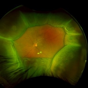
Optos Color Montage of Uveal Effusion Syndrome
Photographer: Drew Mitchell, OCT-C
Imaging device: Optos California
Condition/keywords: color photo, montage, OPTOS, uveal effusion
-
 Uveal Effusion Syndrome
Jan 7 2025 by Drew Mitchell
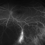
Optos FA Late of Uveal Effusion Syndrome
Photographer: Drew Mitchell, OCT-C
Imaging device: Optos California
Condition/keywords: FA late phase, Optos, uveal effusion
-
 Uveal Effusion Syndrome
Jan 7 2025 by Drew Mitchell
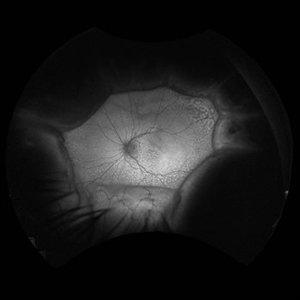
Fundus Autofluorescence Montage of Uveal Effusion Syndrome.
Photographer: Drew Mitchel, OCT-C
Imaging device: Optos California
Condition/keywords: montage, Optos, uveal effusion
-
 Uveal Effusion Syndrome in a Nanophthalmic Eye
Apr 3 2025 by Gustavo Uriel Fonseca Aguirre
B-mode ultrasonography of a nanophthalmic eye reveals diffuse choroidal and scleral thickening, annular ciliochoroidal detachment, and sub-Tenon fluid accumulation.
Photographer: Gustavo U. Fonseca Aguirre, Hospital Conde de Valenciana, Ciudad de México
Condition/keywords: nanophthalmos, uveal effusion syndrome
-
 Uveal Effusion Syndrome in a Nanophthalmic Eye
Apr 3 2025 by Gustavo Uriel Fonseca Aguirre
Ultrasound biomicroscopy longitudinal section of a nanophthalmic eye demonstrating a shallow anterior chamber, angle closure, ciliary body edema, and supraciliary and sub-Tenon fluid accumulation.
Photographer: Gustavo U. Fonseca Aguirre, Hospital Conde de Valenciana, Ciudad de México
Condition/keywords: nanophthalmos, uveal effusion syndrome
-
 Uveal Effusion Syndrome in a Nanophthalmic Eye
Apr 3 2025 by Gustavo Uriel Fonseca Aguirre
Combined B-mode ultrasound with A-mode vector analysis in a nanophthalmic eye (axial length: 14.49 mm).
Photographer: Gustavo U. Fonseca Aguirre, Hospital Conde de Valenciana, Ciudad de México
Condition/keywords: nanophthalmos, uveal effusion syndrome
-
360 Choroidal Detachment in Uveal Effusion Syndrome
Apr 11 2025 by Siri Uppuluri
Fundus photograph of a phakic left eye in an 82-year-old man demonstrating 360 choroidal detachment secondary to uveal effusion syndrome. He underwent sclerectomy and drainage of the choroidal effusions with resolution after surgical intervention.
Photographer: Siri Uppuluri, MD; Rutgers New Jersey Medical School
Condition/keywords: choroidal detachment
-
 Idiopathic Uveal Effusion Syndrome
Aug 24 2012 by John S. King, MD
Resolved on own in a few months; was taking PO NSAIDS.
Photographer: Kristin Konecki, OcuSight Eye Care Center, Rochester, NY
Condition/keywords: idiopathic uveal effusion syndrome, NSAIDs
-
 Idiopathic Uveal Effusion Syndrome
Aug 22 2024 by Jordyn Beckman
61 year old male with Idiopathic Uveal Effusion Syndrome with starry night appearance on fluorescein. 3 weeks s/p single external drainage retinotomy and 9 weeks of oral pred with recurrent choroidal effusions. Has since returned to surgery for secondary drainage retinotomy; subretinal fluid remain persistent.
Photographer: Jordyn Beckman
Imaging device: Optos California
Condition/keywords: chorioretinitis, Choroidal, exudative detachment, window defect
-
 Idiopathic Uveal Effusion Syndrome
Jun 13 2023 by Ahmad B. Tarabishy, MD
66 year old male presented with a 4 month vision of painless decreased vision in the left eye. Clinical findings consistent with idiopathic uveal effusion syndrome. Fundus photography shows 360 degree choroidal elevation with dependent inferior subretinal fluid.
Photographer: Dr. Angela Rico, Retina Specialists of Tampa
Imaging device: Idiopathic Uveal Effusion Syndrome
Condition/keywords: idiopathic uveal effusion syndrome, uveal effusion
-
 Idiopathic Uveal Effusion Syndrome
Jun 13 2023 by Ahmad B. Tarabishy, MD
66 year old male presented with a 4 month vision of painless decreased vision in the left eye. Clinical findings consistent with idiopathic uveal effusion syndrome. Fundus autofluorescence imaging shows presence of leopard spotted pigmentation.
Photographer: Dr. Angela Rico, Retina Specialists of Tampa
Condition/keywords: idiopathic uveal effusion syndrome, uveal effusion
-
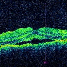 Idiopathic Uveal Effusion Syndrome
Jun 13 2023 by Ahmad B. Tarabishy, MD
66 year old male presented with a 4 month vision of painless decreased vision in the left eye. Clinical findings consistent with idiopathic uveal effusion syndrome. Macular OCT shows presence of subretinal fluid.
Photographer: Dr. Angela Rico, Retina Specialists of Tampa
Condition/keywords: idiopathic uveal effusion syndrome, uveal effusion
 Loading…