Search results (35 results)
-
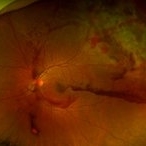 Blunt Ocular Trauma with Commotio Retinae
Nov 5 2019 by Nichole Lewis
11-year-old male with blunt ocular trauma from a soccer ball. Commotio Retinae, retinal hemorrhages, vitreous hemorrhage, multiple retinal tears and a traumatic macular hole. VA 20/70.
Photographer: Nichole Lewis
Imaging device: Optos
Condition/keywords: blunt trauma, commotio retinae, retinal hemorrhage, retinal tear, traumatic macular hole, vitreous hemorrhage
-
 Choroidal Rupture, Subretinal and Vitreous Hemorrhage Secondary to Blunt Trauma
Dec 29 2012 by Humberto Ruiz-Garcia, MD
SD-OCT obtained at 72 hours which shows neurosensory macular detachment and severe thinning (impending macular hole).
Photographer: Humberto Ruiz-Garcia
Imaging device: Cirrus HD OCT
Condition/keywords: traumatic macular hole
-
 Fundus Photograph (Follow-up)
May 8 2021 by Jazli Tan
Follow-up fundoscopic examination revealed decreased subretinal blood and oedema at one week post injury, with persistent macular hole.
Condition/keywords: traumatic macular hole
-
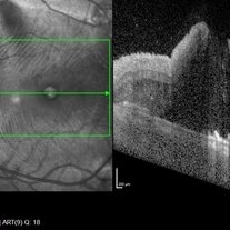
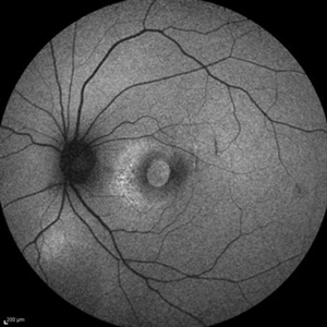
 OCT - Traumatic Full Thickness Macular Hole
Feb 6 2019 by awaneesh m upadhyay, MBBS, DNB
Right eye OCT image of an 8-year-old boy shows full thickness macular hole following blunt trauma of 1 week duration.
Photographer: Awaneesh Upadhyay
Imaging device: HEILDERBERG - HRA
Condition/keywords: traumatic macular hole
-
 Optical Coherence Tomography
May 8 2021 by Jazli Tan
Optical Coherence Tomography (OCT) of a 31-year-old man with a traumatic macular hole from a badminton racket injury. OCT with enhanced depth imaging showed full thickness macular hole with subretinal fluid.
Condition/keywords: traumatic macular hole
-
---thumb.jpg/image-square;max$300,300.ImageHandler) Traumatic Choroidal Rupture and Macular Hole
Traumatic Choroidal Rupture and Macular Hole
Jan 1 2013 by John T. Thompson, MD
Traumatic macular hole and choroidal rupture following blunt trauma to globe.
Condition/keywords: blunt trauma, choroidal rupture, traumatic macular hole
-
 Traumatic Macular Hole
Aug 23 2012 by Gabriela Lopezcarasa Hernandez, MD
12-year-old boy with blunt trauma in right eye and central scotoma.
Photographer: Gabriela Lopezcarasa Hernandez, Hospital Angeles Lomas
Imaging device: ZEISS F4
Condition/keywords: blunt trauma, central scotoma, macular hole
-
---thumb.jpg/image-square;max$300,300.ImageHandler) Traumatic Macular Hole
Traumatic Macular Hole
Mar 9 2013 by Gabriela Lopezcarasa Hernandez, MD
Traumatic macular hole.
Photographer: Gabriela Lopezcarasa Hdz. MD. Hospital Angeles Lomas
Imaging device: zEISS FF4
Condition/keywords: macular hole
-
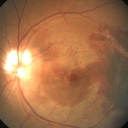
 Traumatic Macular Hole
Aug 23 2012 by Gerardo Garcia-Aguirre, MD
Fundus photograph of a large macular hole with an area of pigment migration secondary to blunt trauma.
Photographer: Noemí Hernández, Asociación para Evitar la Ceguera en México
Imaging device: Zeiss FF4
Condition/keywords: deformity, macular hole
-
 Traumatic Macular Hole
Mar 27 2019 by Gary R. Cook, MD, FACS
14-year-old white male with a traumatic macular hole OS 23 days post hockey puck injury; V.A.= 20/200.
Imaging device: Topcon VT-50
Condition/keywords: full thickness macular hole, macular hole, trauma, traumatic macular hole
-
 Traumatic Macular Hole
Mar 27 2019 by Gary R. Cook, MD, FACS
7-year-old white male with a traumatic macular hole and secondary epiretinal membrane formation OS; hit in the eye with a rock; V.A.= counting fingers.
Imaging device: Topcon VT-50
Condition/keywords: epiretinal membrane formation, full thickness macular hole, macular hole, traumatic macular hole
-
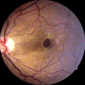
 Traumatic macular hole
Dec 19 2012 by Eric A. Postel, MD
Color fundus photograph of a young male with a traumatic macular hole
Condition/keywords: blunt trauma, macular hole
-
 Traumatic Macular Hole
Aug 18 2024 by Hemanth Murthy, MBBS, MD, FASRS
A 24 year old male patient presented with history of injury at workplace followed by loss of vision. He had a intraocular foreign body with a large traumatic macular hole. Patient was operated and the intraocular foreign body was removed. The hole was too large to close by ILM so an AMG graft was used. Patient regained 20/120 vision.
Photographer: Mr Veda Vyas
Imaging device: Optos Daytona
Condition/keywords: human amniotic graft, traumatic macular hole
-
 Traumatic Macular Hole
Aug 18 2024 by Hemanth Murthy, MBBS, MD, FASRS
A 24 year old male patient presented with history of injury at workplace followed by loss of vision. He had a intraocular foreign body with a large traumatic macular hole. Patient was operated and the intraocular foreign body was removed. The hole was too large to close by ILM so an AMG graft was used. Patient regained 20/120 vision.
Photographer: Mr Veda Vyas
Imaging device: Topcon Triton
Condition/keywords: human amniotic graft, traumatic macular hole
-
 Traumatic Macular Hole
Sep 14 2014 by Mehul A Shah
15-year-old boy presented with cricket ball injury.
Photographer: Drashti Netralaya,Dahod
Imaging device: Zeiss ff450
Condition/keywords: traumatic macular hole
-
 Traumatic Macular Hole
Sep 14 2014 by Mehul A Shah
17-year-old presented with blunt trauma.
Photographer: Drashti Netralaya,Dahod
Imaging device: Zeiss ff450
Condition/keywords: traumatic macular hole
-
 Traumatic Macular Hole
Sep 14 2014 by Mehul A Shah
20-year-old presented with cricket ball injury.
Photographer: Drashti Netralaya,Dahod
Imaging device: Zeiss ff450
Condition/keywords: traumatic macular hole
-
 Traumatic Macular Hole
Oct 2 2017 by Mehul A Shah
A 43-year-old male presented with history of blunt trauma before 6 months. Clinical picture was presented to us.
Photographer: Mehul Shah
Condition/keywords: traumatic macular hole
-
 Traumatic macular hole
Nov 4 2022 by rodrigo torres
Traumatic macular hole
Photographer: Rodrigo Torres
Condition/keywords: choroidal rupture, macular hole, Trauma
-
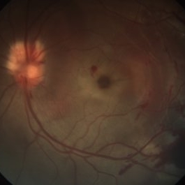 Traumatic Macular Hole
May 8 2021 by Jazli Tan
Fundus photograph of a 31-year-old man with a traumatic macular hole from a badminton racket injury. Fundoscopic examination revealed macular hole with associated sub-retinal, sub-retinal pigment epithelium, and sub-hyaloid bleed temporal to macula. Myelinated nerve fibre layer of the optic disc was noted superiorly with splinter hemorrhages.
Condition/keywords: traumatic macular hole
-
 Traumatic Macular Hole
Feb 6 2019 by awaneesh m upadhyay, MBBS, DNB
Fundus photograph of an 8-year-old boy with vision of <20/200 in OD following blunt trauma of 10 days duration show macular hole with Berlin's edema.
Photographer: Dr. Awaneesh Upadhyay
Condition/keywords: Berlin's edema
-
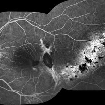 Traumatic Macular Hole And Bruch Membrane Rupture
Jan 22 2021 by Renata Garcia Franco, Md
FA shows hypofluorescence in early frames due to a break in choriocapillaris and choroidal vessels at the rupture site with staning at late phases.
Photographer: Fatima Hernandez, Instituto de la Retina del Bajio SC
Imaging device: Zeiss
Condition/keywords: traumatic macular hole
-
 Traumatic Macular Hole And Bruch Membrane Rupture
Jan 22 2021 by Renata Garcia Franco, Md
Male with history of ocular blunt injury, full-thickness macular hole, RPE changes and Bruch membrane rupture.
Photographer: Fatima Hernandez, Instituto de la Retina del Bajio SC
Imaging device: Zeiss
Condition/keywords: traumatic macular hole
-
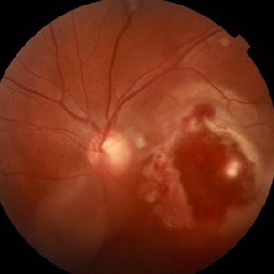
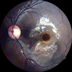
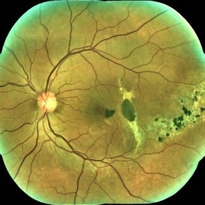
 Traumatic Macular Hole and Choroidal Ruptures
Mar 15 2017 by Hamid Ahmadieh, MD
Fundus autofluorescence ( FAF) image of the left eye of a 30 -year-old woman with a recent history of closed eye injury leading to a large traumatic macular hole and two concentric choroidal ruptures.
Photographer: Solmaz Shahmohammad, Negah Eye Center, Tehran,Iran
Condition/keywords: choroidal rupture, fundus autofluorescence (FAF), traumatic macular hole
-
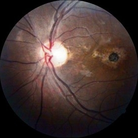
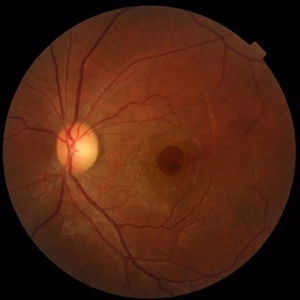 Traumatic Macular Hole and Choroidal Ruptures
Mar 15 2017 by Hamid Ahmadieh, MD
Color fundus photograph of the left eye of a 30 -year-old woman with a history of closed eye injury leading to a large traumatic macular hole and two concentric choroidal ruptures.
Photographer: Solmaz Shahmohammad, Negah Eye Center, Tehran,Iran
Condition/keywords: choroidal rupture, color fundus photograph, traumatic macular hole
 Loading…