Search results (39 results)
-
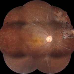
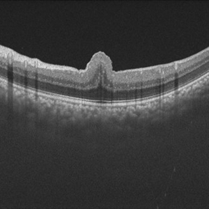
 Astrocytoma OCT
Jan 9 2018 by Sidra Zafar
OCT imaging of retinal astrocytoma in a female child with known diagnosis of tuberous sclerosis. A cystic pocket can be observed.
Imaging device: Swept Source
Condition/keywords: tuberous sclerosis
-
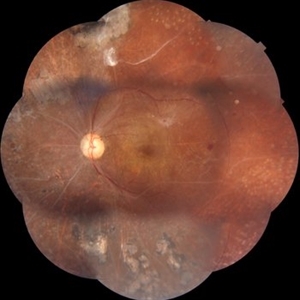
 Astrocytoma OCT
Jan 9 2018 by Sidra Zafar
Swept source OCT imaging of retinal astrocytoma in a female child with known diagnosis of tuberous sclerosis.
Imaging device: Swept Source
Condition/keywords: astrocytoma, optical coherence tomography (OCT), tuberous sclerosis
-
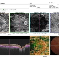
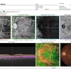
 Multimodal Imaging for Differentiating Unilateral Pseudo Optic Disc Swelling(Buried Drusen) From True Optic Disc Swelling
Feb 7 2024 by Fawwaz F Al Mamoori, MD, Medical Retina Consultant
27-year-old male, medically free, presented with left unilateral optic disc swelling. BCVA=1.0(OU), color vision, and contrast sensitivity were normal (OU)with no RAPD in the left eye. Swept Source OCT: showed elevated left optic disc with hyporeflective mass (Fig-1 B). Enface OCT: Showed left peripapillary multiple ovoid mass lesions(drusen) (Fig-2 d, Fig3 F). FAF: of the left eye showed superonasal hyper autofluorescent drusenoid lesions)(Fig3 E). Orbital MRI with contrast was requested to exclude any compressive lesions like tumors(menigioma)or inflammatory lesions like granuloma(sarcoid granuloma). orbital MRI result was normal.
Photographer: Hana.S.Owais
Imaging device: TRITON(TOPCON,Swept Source OCT)
Condition/keywords: fundus autofluorescence (FAF), multimodal imaging, OCT EN FACE, optic disc drusen, optic disc edema, swept source
-
 OCT of a sub-internal limiting membrane hemorrhage in Valsalva retinopathy
Jul 29 2022 by JORGE SOBERANES
Optical coherence tomography of a 70-year-old man with a sub-internal limiting membrane due to Valsalva retinopathy
Photographer: Jorge I. Soberanes, Asociación para Evitar la Ceguera en México.
Imaging device: PLEX Elite 9000, Zeiss
Condition/keywords: OCT, sub-inner limiting membrane hemorrhage, swept source, valsalva retinopathy
-
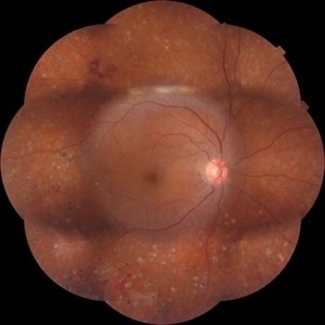
 Proliferative Diabetic Retinopathy
Mar 1 2021 by Avris Romario Diparaja Siahaan
Fundus photograph (montage photography) of a 57-year-old woman with proliferative diabetic retinopathy in her both eyes.
Photographer: Nanda Lessi Hafni Eka Putri, MD (Ophthalmologist) & Ryan Mishbahuddin (Nurse), Ciawi General Hospital (Rumah Sakit Umum Daerah Ciawi)
Imaging device: DRI OCT Triton Plus
Condition/keywords: fundus photograph, montage, optical coherence tomography (OCT), swept source, wide angle imaging
-
 Proliferative Diabetic Retinopathy
Mar 1 2021 by Avris Romario Diparaja Siahaan
Swept Source OCT angiography (montage photography) of a 57-year-old woman with proliferative diabetic retinopathy in her both eyes.
Photographer: Nanda Lessi Hafni Eka Putri, MD (Ophthalmologist) & Ryan Mishbahuddin (Nurse), Ciawi General Hospital (Rumah Sakit Umum Daerah Ciawi)
Imaging device: DRI OCT Triton Plus
Condition/keywords: fundus photograph, montage, optical coherence tomography (OCT), swept source, wide angle imaging
-
 Proliferative Diabetic Retinopathy
Mar 1 2021 by Avris Romario Diparaja Siahaan
Swept source OCT angiography (montage photography) of a 57-year-old woman with proliferative diabetic retinopathy in her both eyes.
Photographer: Nanda Lessi Hafni Eka Putri, MD (Ophthalmologist) & Ryan Mishbahuddin (Nurse), Ciawi General Hospital (Rumah Sakit Umum Daerah Ciawi)
Imaging device: DRI OCT Triton Plus
Condition/keywords: fundus photograph, montage, optical coherence tomography (OCT), swept source, wide angle imaging
-
 Proliferative Diabetic Retinopathy
Mar 1 2021 by Avris Romario Diparaja Siahaan
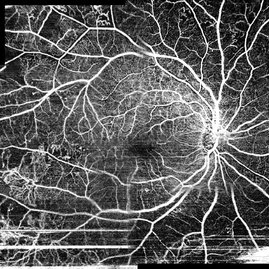
Swept source OCT angiography (12.0 mm X 12.0 mm) of a 57-year-old woman with proliferative diabetic retinopathy in her both eyes.
Photographer: Nanda Lessi Hafni Eka Putri, MD (Ophthalmologist) & Ryan Mishbahuddin (Nurse), Ciawi General Hospital (Rumah Sakit Umum Daerah Ciawi)
Imaging device: DRI OCT Triton Plus
Condition/keywords: fundus photograph, montage, optical coherence tomography (OCT), swept source, wide angle imaging
-
 Proliferative Diabetic Retinopathy
Mar 1 2021 by Avris Romario Diparaja Siahaan
Swept source OCT angiography (12.0 mm X 12.0 mm) of a 57-year-old woman with proliferative diabetic retinopathy in her both eyes.
Photographer: Nanda Lessi Hafni Eka Putri, MD (Ophthalmologist) & Ryan Mishbahuddin (Nurse), Ciawi General Hospital (Rumah Sakit Umum Daerah Ciawi)
Imaging device: DRI OCT Triton Plus
Condition/keywords: fundus photograph, montage, optical coherence tomography (OCT), swept source, wide angle imaging
-
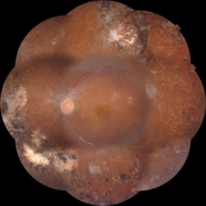
 Proliferative Diabetic Retinopathy
Mar 1 2021 by Avris Romario Diparaja Siahaan
Fundus photograph (montage) of a 62-year-old woman with proliferative diabetic retinopathy in her both eyes.
Photographer: Nanda Lessi Hafni Eka Putri, MD (Ophthalmologist) & Ryan Mishbahuddin (Nurse), Ciawi General Hospital (Rumah Sakit Umum Daerah Ciawi)
Imaging device: DRI OCT Triton Plus
Condition/keywords: fundus photograph, montage, optical coherence tomography (OCT), swept source, wide angle imaging
-
 Proliferative Diabetic Retinopathy
Mar 1 2021 by Avris Romario Diparaja Siahaan
Swept source OCT angiography (montage photography) of a 62-year-old woman with proliferative diabetic retinopathy in her both eyes.
Photographer: Nanda Lessi Hafni Eka Putri, MD (Ophthalmologist) & Ryan Mishbahuddin (Nurse), Ciawi General Hospital (Rumah Sakit Umum Daerah Ciawi)
Imaging device: DRI OCT Triton Plus
Condition/keywords: fundus photograph, montage, optical coherence tomography (OCT), swept source, wide angle imaging
-
 Proliferative Diabetic Retinopathy
Mar 1 2021 by Avris Romario Diparaja Siahaan
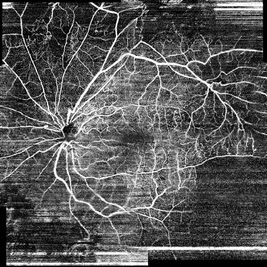
Swept Source OCT angiography (9.0 mm X 9.0 mm) of a 62-year-old woman with proliferative diabetic retinopathy in her both eyes.
Photographer: Nanda Lessi Hafni Eka Putri, MD (Ophthalmologist) & Ryan Mishbahuddin (Nurse), Ciawi General Hospital (Rumah Sakit Umum Daerah Ciawi)
Imaging device: DRI OCT Triton Plus
Condition/keywords: fundus photograph, montage, optical coherence tomography (OCT), swept source, wide angle imaging
-
 Proliferative Diabetic Retinopathy
Mar 1 2021 by Avris Romario Diparaja Siahaan
Swept Source OCT angiography (9.0 mm X 9.0 mm) of a 62-year-old woman with proliferative diabetic retinopathy in her both eyes.
Photographer: Nanda Lessi Hafni Eka Putri, MD (Ophthalmologist) & Ryan Mishbahuddin (Nurse), Ciawi General Hospital (Rumah Sakit Umum Daerah Ciawi)
Imaging device: DRI OCT Triton Plus
Condition/keywords: fundus photograph, montage, optical coherence tomography (OCT), swept source, wide angle imaging
-
 Vitreous Hemorrhage (Floater Storm)
Jan 3 2025 by Drew Mitchell
27mm line scan on the Optos Silverstone of a new Vitreous Hemorrhage.
Photographer: Drew Mitchell, OCT-C
Imaging device: Optos Silverstone
Condition/keywords: Optos, swept source, Vitreous hemorrhage
-
 Whole Eye OCT
Jan 4 2019 by Netan Choudhry, MD, FRCS(C) FASRS
Swept-Source OCT montage of a 45-year-old male with Alports disease and posterior subcapsular cataract.
Photographer: John Golding BA, Vitreous Retina Macula Specialists of Toronto
Imaging device: Topcon DRI Triton
Condition/keywords: Alports disease, optical coherence tomography (OCT), swept source
-
 Diabetic Retinopathy
Sep 2 2021 by Avris Romario Diparaja Siahaan
Swept source OCT angiography (montage photography) and fundus photography of a 61-year-old woman with proliferative diabetic retinopathy in her right eye.
Photographer: Nanda Lessi Hafni Eka Putri, MD (Ophthalmologist) & Ryan Mishbahuddin (Nurse), Ciawi General Hospital (Rumah Sakit Umum Daerah Ciawi)
Imaging device: DRI OCT Triton Plus (Topcon)
Condition/keywords: diabetic retinopathy, fundus photograph, optical coherence tomography (OCT)
-
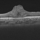
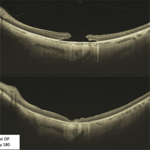 Large Macular Hole: Pre-op and Post-op
Jan 4 2022 by Thirumalesh Mochi Basavaraj, MD
Swept source OCT of a 65-yea-old patient with a large macular hole before and after surgery.
Photographer: Puttaswamy Narayana Nethralaya
Imaging device: Topcon Dri Triton
Condition/keywords: macular hole
-
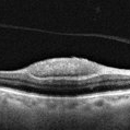
 Macular Hole
Jan 3 2022 by Thirumalesh Mochi Basavaraj, MD
Swept source OCT of a 65-year-old patient with posterior hyaloid separation and a large macular hole with undermined edges.
Photographer: Puttaswamy Narayana Nethralaya Bangalore
Imaging device: Topcon Dri Triton
Condition/keywords: large macular hole, posterior cyclitis hyaloid separation, swept source OCT
-
 OCT en face of a 360 retinotomy for closed funnel combined retinal detachment
Jan 1 2023 by Malek Yassine, MD
Swept source OCT en face at the silicon oil - Retina Interface shows droplets of SO emulsification around the fovea and at the superior arcade, with some inferior striae corresponding to ERM formation
Imaging device: Topcon Triton DRI-OCT
Condition/keywords: oct en face
-
 OCT en face of a 360 retinotomy for closed funnel combined retinal detachment
Jan 1 2023 by Malek Yassine, MD
Swept Source OCT en face at deep capillary plexus, shows foveal and parafoveal intraretinal cysts corresponding to macular edema under silicon oil
Imaging device: Topcon Triton DRI-OCT
Condition/keywords: combined retinal detachment, OCT EN FACE
-
 Resolving of Refractory Subretinal Fluid Post Faricimab Injection
Nov 7 2022 by Fawwaz F Al Mamoori, MD, Medical Retina Consultant
A case of Rt eye wet-AMD treated over 2 years with a Q8weeks aflibercebt injection with refractory SRF less than 200 um that shifted recently to Faricimab.1 month post Faricimab OCT showed a complete drying out of refractory subretinal Fluid (SRF)
Photographer: Mohammed Rabaa
Imaging device: Swept Source OCT (TRITON) Topcon
Condition/keywords: subretinal fluid
-
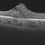
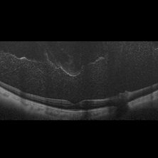
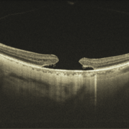
 Retinal Fold in Posterior Microphthalmos
Mar 1 2025 by Hemanth Murthy, MBBS, MD, FASRS
Swept source OCT image of Right eye of 34 year male patient with high hypermetropia(+14). BCVA 20/20 in right eye and 20/60 in left eye. Anterior segment was normal. There is loss of foveal pit with omega shaped elevation of inner retinal layers.
Photographer: Mr Veda Vyas
Condition/keywords: posterior microphthalmos
 Loading…