Search results (74 results)
-
 Angioid Streaks
Mar 11 2014 by Andrew M Hendrick, MD
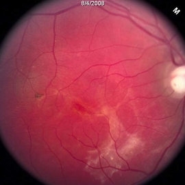
Fundus photography of the left eye of a 50-year-old African American male with a remote history of minor trauma in the contralateral eye.
Condition/keywords: angioid streaks, subretinal fibrosis
-
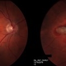
 Angioid Streaks With CNV
Mar 11 2014 by Andrew M Hendrick, MD
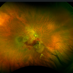
Fundus photography of the right eye of a 50-year-old African American male with a remote history of minor trauma. Serial anti-VEGF injections failed to improve the subfoveal CNV and his condition is now being observed.
Photographer: Jannah Dobbs
Condition/keywords: angioid streaks, subretinal fibrosis
-
 Central Serous Chorioretinopathy (CSC)
Oct 16 2012 by S. Natarajan, MD, FASRS, FRCS (GLASGOW) , FICO, D.Sc, FELA
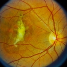
Middle-aged male came with small PED 4 months back; now this has progressed to a larger PED with SRF underneath the fovea.
Photographer: Prof. Dr. S. Natarajan
Condition/keywords: central serous chorioretinopathy (CSCR), central serous retinopathy (CSR), pigment epithelial detachment (PED), subretinal fibrosis
-
 Chorioretinal Scars with Subretinal Fibrosis and an old Retinal Detachment
May 3 2018 by Nichole Lewis
Chorioretinal scars with subretinal fibrosis and an old retinal detachment.
Photographer: Nichole Lewis
Condition/keywords: chorioretinal scar, chronic retinal detachment, subretinal fibrosis
-
 Diffuse Choroidal Hemangioma
Nov 7 2012 by Rajiv Anand, MD, FRCS, FASRS
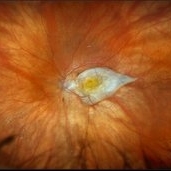
Fundus photo shows classic 'tomato-ketchup' red appearance of diffuse hemangioma. Due to chronic SRF , there is subretinal fibrosis.
Condition/keywords: subretinal fibrosis
-
 Inferior Rhegmatogenous Retinal Detachment with Subretinal Fibrosis
Aug 23 2012 by Gabriela Lopezcarasa Hernandez, MD
Asymptomatic 25-year-old woman with high myopia.
Photographer: Gabriela Lopezcarasa Hernandez, Hospital Angeles Lomas
Imaging device: FF4
Condition/keywords: high myopia, subretinal fibrosis
-
 multifocal choroiditis
Feb 14 2013 by From the Collections of Thomas M. Aaberg, MD and Thomas M. Aaberg Jr., MD
color fundus photos showing healed chorioretinal scars, pigment deposition, and subretinal fibrosis consistent with regressed multifocal choroiditis
Condition/keywords: multifocal choroiditis, posterior segment inflammation, subretinal fibrosis, white dot syndrome
-
 Neovascular Age-Related Macular Degeneration (1)
Apr 28 2021 by Ambar Faridi, MD
80-year-old woman with neovascular age-related macular degeneration with large subretinal hemorrhage, hemorrhagic PED, and vascular lipid exudation.
Photographer: Jennifer Tu-Bui, VA Portland Health Care System
Condition/keywords: subretinal fibrosis, subretinal hemorrhage
-
 Peculiar Acute Subretinal Fibrosis
Feb 24 2015 by David Callanan, MD
Peculiar acute subretinal fibrosis.
Condition/keywords: subretinal fibrosis
-
 Peculiar Acute Subretinal Fibrosis
Feb 24 2015 by David Callanan, MD
Peculiar acute subretinal fibrosis.
Condition/keywords: subretinal fibrosis
-
 Peculiar Acute Subretinal Fibrosis
Feb 24 2015 by David Callanan, MD
Peculiar acute subretinal fibrosis.
Condition/keywords: subretinal fibrosis
-
 Peculiar Acute Subretinal Fibrosis
Feb 24 2015 by David Callanan, MD
Peculiar acute subretinal fibrosis.
Condition/keywords: subretinal fibrosis
-
 Peculiar Acute Subretinal Fibrosis
Feb 24 2015 by David Callanan, MD
Peculiar acute subretinal fibrosis.
Condition/keywords: subretinal fibrosis
-
 Subretinal Fibrosis
Feb 20 2015 by H. Michael Lambert, MD
Posterior pole with possible subretinal fibrosis.
Condition/keywords: subretinal fibrosis
-
 Subretinal Fibrosis
Feb 20 2015 by H. Michael Lambert, MD
Posterior pole with possible subretinal fibrosis.
Condition/keywords: subretinal fibrosis
-
 Subretinal fibrosis
Sep 14 2023 by Ben Serar
Fundus photograph of LE showing a scarred lesion at the macula, with sub retinal fibrosis.
Condition/keywords: macular scar, Subretinal fibrosis
-
 Subretinal fibrosis
Sep 12 2023 by Ben Serar
Fundus photograph of RE showing scarring at the macula with subretinal fibrosis
Condition/keywords: Subretinal fibrosis
-
 Subretinal Fibrosis
Jan 14 2025 by Kimberly Wakester
Fundus photograph of an 86-year-old woman with the end stage of Age-related Macular Degeneration in the left eye. Patient went unseen for 3-4 years prior to establishing care at our practice. Due to the significant amount of subretinal fibrosis, treatment was not recommended due to limited visual recovery. Patient was advised of monocular vision and the importance of follow up care.
Photographer: Kimberly Wakester, COA
Imaging device: Optos California
Condition/keywords: AMD, subretinal fibrosis
-
 Subretinal Fibrosis and Uveitis Syndrome
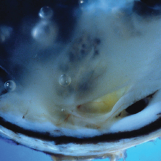
May 18 2020 by McGill University Health Centre
Uveitis syndrome is a rare posterior uveitis that usually begins as a multifocal choroiditis and then progresses to subretinal fibrosis. Recurrences are not uncommon and the visual prognosis is generally poor. In this enucleation specimen, a thickened choroid is clearly observed (arrow). The retina is detached and a fibrovascular subretinal membrane is present (arrowhead).
Condition/keywords: subretinal fibrosis, uveitis
-
 Subretinal Thickening and Subretinal Hemorrhage – Stereo Color Fundus Photograph
Mar 9 2017 by James B. Soque, CRA, OCT-C, COA, FOPS
Color fundus stereo photograph of a 52-year-old white male with VA loss to 20/200 of unknown etiology. Dilated fundus examination of the right eye reveals a fibrotic scar with subretinal thickening and subretinal hemorrhage.
Photographer: James B Soque, CRA, OCT-C, COA
Imaging device: Topcon TRC 50 DX, MERGE Software
Condition/keywords: blood, color fundus photograph, color photo, stereo pair, subretinal blood, subretinal fibrosis, subretinal thickening
-
 VKH Pseudotumor – Chronic Subretinal Fibrosis
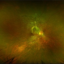
May 11 2025 by Felipe Murati
Ultra-widefield fundus image from a 36-year-old woman with chronic VKH syndrome showing a pseudotumor-like subretinal fibrotic lesion in the right eye. The lesion developed after multiple relapses and remained stable over a 1-year follow-up with immunosuppressive treatment including prednisone, mycophenolate mofetil, and adalimumab. No active choroiditis or exudative detachment was observed. Multimodal imaging was essential for disease monitoring.
Photographer: Felipe A. Murati, MD, University of Arizona
Imaging device: Optos California ultra-widefield retinal imaging system, single-capture, color fundus modality.
Condition/keywords: adalimumab, chronic inflammation, granulomatous uveitis, OCT, Optos ultra-widefield imaging, pseudotumor, subretinal fibrosis, VKH, Vogt-Koyanagi-Harada
-
 VKH Pseudotumor – Fluorescein Angiography
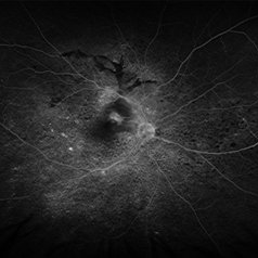
May 11 2025 by Felipe Murati
Fluorescein angiography image from a 36-year-old woman with chronic Vogt-Koyanagi-Harada (VKH) syndrome showing a pseudotumor-like lesion with late-phase staining and no active leakage. The image highlights subretinal fibrosis in the right eye, stable under long-term immunosuppressive therapy with mycophenolate mofetil and adalimumab. No signs of active choroiditis are present, confirming a quiescent phase.
Photographer: Felipe A. Murati, MD, University of Arizona
Imaging device: Optos California, fluorescein angiography modality
Condition/keywords: choroiditis, Fluorescein angiography, granulomatous uveitis, Optos FA, pseudotumor, subretinal fibrosis, VKH, Vogt-Koyanagi-Harada
-
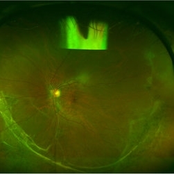 Wagner Syndrome
Aug 1 2017 by Eitae Kim, MD
Ulltra wide field fundus photograph of 19-year-old male with Wagner syndrome which shows peripheral subretinal fibrosis and pigmentary degeneration.
Photographer: Eitae Kim, BOIM retinal center, Pureun eye hospital
Condition/keywords: subretinal fibrosis, ultra-wide field imaging, Wagner disease
-
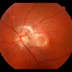 Subretinal Fibrosis (PPCNVM and POHS) OS
Sep 18 2019 by John S. King, MD
57-year-old white male with history of PPCNVM OS and POHS OU here for a routine visit. History of avastin in 2014, and stable since then. Va OS 20/20. PP scar with macular subretinal fibrosis. No heme or exudates. CR spot supero-nasally.
Photographer: Shelly Blair
Imaging device: Topcon 50
Condition/keywords: choroidal neovascular membrane (CNVM), ocular histoplasmosis syndrome (OHS), peripapillary choroidal neovascularization (PPCNVM), presumed ocular histoplasmosis syndrome (POHS)
-
 Angioid streaks - PXE
Jan 11 2013 by Alex P. Hunyor, MD
Pseudoxanthoma elasticum with angioid streaks, left eye - note subretinal fibrosis adjacent to disc.
Condition/keywords: angioid streaks, pseudoxanthoma elasticum (PXE)
 Loading…