Search results (111 results)
-
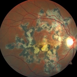
 FA Serpiginous Choroidal Atrophy & CNV OS
May 15 2024 by Angela Rico
33 year-old female. Negative TB. No H/O immunosuppression. OD: 5:02 Timer OS: 2:12 Timer
Photographer: Angela Rico M.D.
Condition/keywords: serpiginous choroiditis
-
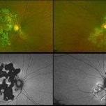
 Fundus Autofluorescense Serpiginious Chorioretinitis
Sep 6 2023 by PUSHPANJALI BADOLE
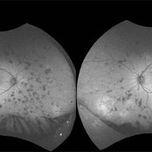
Optos fundus autofluorescense image shows multiple hyper and hypo autofluorescent lesions corresponding to active and healed lesions in the midperipheral retina and periphery suggestive of varied stage presentation of lesions.
Photographer: Hitesh Rawlani, Isha Netralaya, Kalyan.
Condition/keywords: serpiginous choroiditis
-
 Macular Serpiginous Choroidopathy
Sep 27 2012 by Raj K. Maturi, MD
9/11/2012
Photographer: Char Harris
Imaging device: HRA
Condition/keywords: serpiginous choroiditis
-
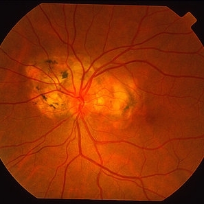
 Serpigenous Choroidopathy in a 68-Year-Old Male
Feb 15 2013 by Roy Schwartz, MD
A 68-year-old healthy male presented with a few years of decreased vision bilaterally. Visual acuity in OD was 1/36 and in OS 20/40. Anterior segments were normal except for bilateral mild nuclear sclerosis and pseudoexfoliation in OS. In the fundus of OD a large atrophy with pigmentary scars were seen in the macula and nasally to the optic disc while OS presented with the same clinical picture but an island of normal appearing retina was seen in the fovea. On fluorscein angiography no leakage was shown. A diagnosis of Serpigenous choroidopathy was made.
Photographer: Galit Yair-Pur
Condition/keywords: macula serpiginous choroidopathy, serpiginous choroiditis
-
 Serpiginious Chorioretinitis
Sep 6 2023 by PUSHPANJALI BADOLE
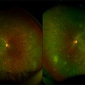
Fundus photograph of an 18 year-old male with bilateral choroiditis. Tuberculosis work-up revealed positive QuantiFERON Gold test and Mantoux test. Patient was started anti-tuberculosis treatment along with oral corticosteroids. Optos fundus photography shows extensive active plus healed lesions with pigmentary change in the midperipheral retina and periphery suggesting varied stage presentation of lesions. There are few hemorrhages in right eye superotemporal retina.
Photographer: Hitesh Rawlani, Isha Netralaya, Kalyan.
Condition/keywords: serpiginous choroiditis
-
 Serpiginous
Aug 29 2012 by F. Ryan Prall, MD
70-year-old female with reactivation of serpiginous choroiditis.
Photographer: Tom Egnatz, Indiana University
Condition/keywords: serpiginous choroiditis
-
 Serpiginous
Aug 29 2012 by F. Ryan Prall, MD
70-year-old female with reactivation of serpiginous choroiditis.
Photographer: Tom Egnatz, Indiana University
Condition/keywords: serpiginous choroiditis
-
 Serpiginous
Aug 29 2012 by F. Ryan Prall, MD
70-year-old female with reactivation of serpiginous choroiditis.
Photographer: Tom Egnatz, Indiana University
Condition/keywords: serpiginous choroiditis
-
 Serpiginous
Aug 29 2012 by F. Ryan Prall, MD
70-year-old female with reactivation of serpiginous choroiditis.
Photographer: Tom Egnatz, Indiana University
Condition/keywords: serpiginous choroiditis
-
 Serpiginous
Aug 29 2012 by F. Ryan Prall, MD
70-year-old female with reactivation of serpiginous choroiditis.
Photographer: Tom Egnatz, Indiana University
Condition/keywords: serpiginous choroiditis
-
 Serpiginous Choroidal Atrophy
-
 Serpiginous Choroidal Atrophy
May 28 2024 by Angela Rico
33 year-old female. Negative For TB or History of Immunosuppression. VA: OD 20/60-2 OS 20/150
Photographer: Angela Rico M.D.
Condition/keywords: macula serpiginous choroidopathy, serpiginous choroiditis
-
 Serpiginous Choroidal Atrophy
May 28 2024 by Angela Rico
33 year-old female. Negative For TB or History of Immunosuppression. VA: OD 20/60-2 OS 20/150
Condition/keywords: macula serpiginous choroidopathy, serpiginous choroiditis
-
 Serpiginous Choroidal Atrophy & CNV OS
May 15 2024 by Angela Rico
33 year-old female. Negative TB. No history of Immunosuppression.
Photographer: Angela Rico M.D.,
Condition/keywords: macula serpiginous choroidopathy, serpiginous choroiditis
-
 Serpiginous Choroiditis
Nov 14 2021 by Maxwell J Wingelaar, MD
An image showing active Serpiginous Choroiditis.
Condition/keywords: serpiginous choroiditis
-
 Serpiginous Choroiditis
Sep 22 2019 by Haider Ali
35-year-old female presented with decrease in vision in her left eye for last 4 days and in right eye for last 8 days. Her right eye was previously involved in a similar episode about 5-6 months ago for which she was treated with oral steroids.
Photographer: Dr Haider Ali Chaudhry, Madinah Teaching Hospital, Faisalabad
Condition/keywords: acute posterior multifocal placoid pigment epitheliopathy (APMPPE), macula serpiginous choroidopathy, posterior uveitis, serpiginous choroiditis, uveitis, white dot lesions, white dot syndrome
-
 Serpiginous Choroiditis
Apr 28 2019 by Nilesh K Kanjani, MD
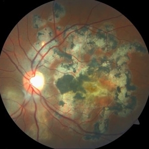
Fundus photo of a young 30-year-old male patient with progressive loss of vision in few weeks.
Photographer: Nilesh Kanjani, Dr Agarwal Eye Hospital, Ahmedabad
Condition/keywords: serpiginous choroiditis
-
 Serpiginous Choroiditis
Sep 16 2012 by Ivan R. Batlle, MD
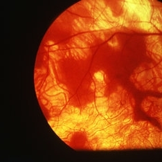
Fluorescein angiogram of 30-year-old female with multiple autoimmune disorders
Condition/keywords: serpiginous choroiditis
-
 Serpiginous Choroiditis
Sep 16 2012 by Ivan R. Batlle, MD
Fluorescein angiogram of 30-year-old female with multiple autoimmune disorders
Condition/keywords: serpiginous choroiditis
-
 Serpiginous Choroiditis
Apr 19 2013 by Brandon G. Busbee, MD
Serpiginous choroiditis.
Photographer: Nichole Lewis - Tennessee Retina - Nashville, TN
Imaging device: Topcon TRC 50-DX
Condition/keywords: serpiginous choroiditis
-
 Serpiginous Choroiditis
Apr 19 2013 by Brandon G. Busbee, MD
Serpiginous choroiditis.
Photographer: Nichole Lewis - Tennessee Retina - Nashville, TN
Imaging device: Topcon TRC 50-DX
Condition/keywords: serpiginous choroiditis
-
 Serpiginous Choroiditis
Feb 25 2013 by Henry J. Kaplan, MD
Serpiginous choroiditis, left eye #2. Active edematous lesion visible moving toward the fovea.
Condition/keywords: serpiginous choroiditis
 Loading…