Search results (66 results)
-
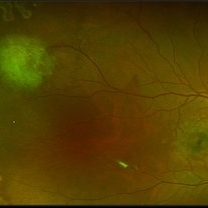
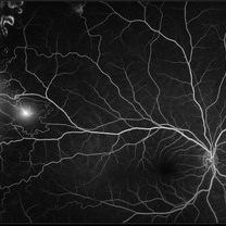
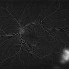
 Active Proliferative Diabetic Retinopathy
Jul 12 2024 by Korey Starkey
Fluorescein angiogram performed on 35 year old female with active proliferative diabetic retinopathy. Patient has peripapillary vascular loop and history of PRP treatment in both eyes. Patients left eye vision measured at Dcc20/200-1 at this visit.
Photographer: Korey Starkey
Imaging device: Optos
Condition/keywords: FLUORESCEIN ANGIOGRAPHY, hyperfluorescence, laser scarring, Optos, proliferative diabetic retinopathy (PDR), sea fan, ultra-wide field imaging, vascular loop
-
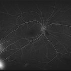
 Eales Disease
Jan 31 2025 by Thirumalesh Mochi Basavaraj, MD
Ultra-wide field image of a 24 year old young healthy adult male with a visible sea fan neovascularization with partial PVD with vitreous and subhyaloid hemorrhage.
Photographer: Puttaswamy
Condition/keywords: Eales disease, sea fan, Ultra-wide field retinal imaging
-
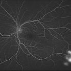
 Eales Disease
Jan 31 2025 by Thirumalesh Mochi Basavaraj, MD
Ultra wide field image of a 24 year-old young healthy adult male with a visible sea fan neovascularization with partial PVD secondary to Scatter LASER photocoagulation with Vitreous and subhyaloid hemorrhage.
Photographer: Puttaswamy N K
Condition/keywords: Eales disease, Neovascularisation elsewhere (NVE), sea fan
-
---thumb.jpg/image-square;max$300,300.ImageHandler) Macro And Microvascular Sea Fan Changes
Macro And Microvascular Sea Fan Changes
-
---thumb.jpg/image-square;max$300,300.ImageHandler) Macro And Microvascular Sea Fan Changes
Macro And Microvascular Sea Fan Changes
-
---thumb.jpg/image-square;max$300,300.ImageHandler) Macro And Microvascular Sea Fan Changes
Macro And Microvascular Sea Fan Changes
-
---thumb.jpg/image-square;max$300,300.ImageHandler) Macro And Microvascular Sea Fan Changes
Macro And Microvascular Sea Fan Changes
-
---thumb.jpg/image-square;max$300,300.ImageHandler) Macro And Microvascular Sea Fan Changes
Macro And Microvascular Sea Fan Changes
-
---thumb.jpg/image-square;max$300,300.ImageHandler) Macro And Microvascular Sea Fan Changes
Macro And Microvascular Sea Fan Changes
-
---thumb.jpg/image-square;max$300,300.ImageHandler) Macro And Microvascular Sea Fan Changes
Macro And Microvascular Sea Fan Changes
-
---thumb.jpg/image-square;max$300,300.ImageHandler) Macro And Microvascular Sea Fan Changes
Macro And Microvascular Sea Fan Changes
-
---thumb.jpg/image-square;max$300,300.ImageHandler) Macro And Microvascular Sea Fan Changes
Macro And Microvascular Sea Fan Changes
-
---thumb.jpg/image-square;max$300,300.ImageHandler) Macro And Microvascular Sea Fan Changes
Macro And Microvascular Sea Fan Changes
-
---thumb.jpg/image-square;max$300,300.ImageHandler) Macro And Microvascular Sea Fan Changes
Macro And Microvascular Sea Fan Changes
-
 Proliferative Diabetic Retinopathy with Temporal Seafan NVE Dragging Retinal Vein
Feb 15 2018 by Kushal S Delhiwala, MBBS, MS, FMRF,FICO, FAICO
58-year-old diabetic male presenting with Bilateral Proliferative diabetic retinopathy and centre involving Diabetic macular edema.Left eye fundus photograph showing large seafan NVE temporal to macula causing upward dragging of inferotemporal retinal vein and arteriovenous anastomosis.
Photographer: Dr Kushal Delhiwala, Netralaya superspeciality eye hospital, Ahmedabad
Imaging device: Zeiss Visucam 500
Condition/keywords: neovascularization elsewhere (NVE), proliferative diabetic retinopathy (PDR), sea fan
-
 Proliferative Sickle Cell Retinopathy
Jan 29 2021 by Olivia Rainey
Ultra-widefield fundus photograph of a 24-year-old female with proliferative sickle cell retinopathy affecting her right eye. He performed scatter PRP OD on 12/2/2020 to nonperfusion in temporal far periphery. The patient's 12/2020 Hb electrophoresis came back showing Hb SC (rather than sickle cell trait). Patient was born at full term, but she reports that her mother used drugs while pregnant with the patient. The patient also mentioned that her niece has full sickle cell disease and her grandmother, mother, and sibling all have condition on the sickle cell spectrum.
Photographer: Olivia Rainey, OCT-C, COA
Imaging device: Optos California
Condition/keywords: fundus photograph, laser photocoagulation, neovascularization (NV), neovascularization elsewhere (NVE), Optos, pseudocolor, sea fan, sickle cell retinopathy
-
 Proliferative Sickle Cell Retinopathy
Jan 29 2021 by Olivia Rainey
Ultra-widefield fluorescein angiogram of a 24-year-old female with proliferative sickle cell retinopathy affecting her right eye. The physician's interpretation of the fluorescein shows seafan neovascularization superotemporally, AV anastomeses, and good peripheral laser. He performed scatter PRP OD on 12/2/2020 to nonperfusion in temporal far periphery. The patient's 12/2020 Hb electrophoresis came back showing Hb SC (rather than sickle cell trait). Patient was born at full term, but she reports that her mother used drugs while pregnant with the patient. The patient also mentioned that her niece has full sickle cell disease and her grandmother, mother, and sibling all have condition on the sickle cell spectrum.
Photographer: Olivia Rainey, OCT-C, COA
Imaging device: Optos California
Condition/keywords: fluorescein angiogram (FA), fluorescein leakage, neovascularization (NV), neovascularization elsewhere (NVE), Optos, sea fan, sickle cell retinopathy
-
 Proliferative Sickle Cell Retinopathy, Color OD
May 23 2018 by Hosam Attia, MD
45-year-old African American, male with sickle cell anemia (SC disease) with arteriolar attenuation, mild venous tortuosity, Sunburst (S) and large, partially auto-infarcted sea fan with fresh heme, OD.
Imaging device: Optos California Ultra-Wide Field Fundus Camera
Condition/keywords: neovascularization elsewhere (NVE), proliferative retinopathy, sea fan, sickle cell, sickle cell retinopathy
-
 Proliferative Sickle Cell Retinopathy, Color OD
May 23 2018 by Hosam Attia, MD
45-year-old African American, male with sickle cell anemia (SC disease) with arteriolar attenuation, mild venous tortuosity, Sunburst (S) and large, partially auto-infarcted Seafan with fresh heme, OD.
Imaging device: Optos California Ultra-Wide Field Fundus Camera
Condition/keywords: neovascularization elsewhere (NVE), proliferative retinopathy, sea fan, sickle cell, sickle cell retinopathy
-
 Proliferative Sickle Cell Retinopathy, Color OS
May 23 2018 by Hosam Attia, MD
45-year-old African American, male with sickle cell anemia (SC disease ) with arteriolar attenuation, mild venous tortuosity, peripheral arterio-venous anastomoses (shown better on red free), multiple small NVEs/ early sea fans (one w/ early auto-infarction) and sunburst (S) - (Not showing very well in photos) OS.
Imaging device: Optos California Ultra-Wide Field Fundus Camera
Condition/keywords: neovascularization elsewhere (NVE), proliferative retinopathy, sea fan, sickle cell, sickle cell retinopathy
-
 Proliferative Sickle Cell Retinopathy, Early phase FA OD
May 23 2018 by Hosam Attia, MD
Fluorescein angiogram photograph of a 45-year-old African American, male with sickle cell anemia (SC disease), depicting extensive peripheral capillary non-perfusion, with early hyperfluorescence over the ischemic retina temporally, with late staining and diffuse leakage consistent with partially auto-infarcted, but active NVE/sea fan OD.
Imaging device: Optos California Ultra-Wide Field Fundus Camera
Condition/keywords: neovascularization elsewhere (NVE), proliferative retinopathy, sea fan, sickle cell, sickle cell retinopathy
-
 Proliferative Sickle Cell Retinopathy, Early phase FA OS
May 23 2018 by Hosam Attia, MD
Fluorescein angiogram photograph of a 45-year-old African American, male with cell anemia (SC disease ), depicting peripheral capillary non-perfusion, with multiple, small area of early to mid phase hyperfluorescence over the ischemic retina temporally, with mild late leakage consistent with active NVEs/ early sea fans OS.
Imaging device: Optos California Ultra-Wide Field Fundus Camera
Condition/keywords: neovascularization elsewhere (NVE), proliferative retinopathy, sea fan, sickle cell, sickle cell retinopathy
-
 Proliferative Sickle Cell Retinopathy, Late FA OS
May 23 2018 by Hosam Attia, MD
Fluorescein angiogram photograph of a 45-year-old African American, male with sickle cell anemia (SC disease), depicting peripheral capillary non-perfusion, with multiple, small area of mild late leakage consistent with active NVEs/ early Seafans OS.
Imaging device: Optos California Ultra-Wide Field Fundus Camera
Condition/keywords: neovascularization elsewhere (NVE), proliferative retinopathy, sea fan, sickle cell, sickle cell retinopathy
-
 Proliferative Sickle Cell Retinopathy, Late phase FA OD
May 23 2018 by Hosam Attia, MD
Fluorescein angiogram photograph of a 45-year-old African American, male with sickle cell anemia (SC disease), depicting extensive peripheral capillary non-perfusion, with late staining and diffuse leakage consistent with partially auto-infarcted, but active NVE/sea fan OD.
Imaging device: Optos California Ultra-Wide Field Fundus Camera
Condition/keywords: neovascularization elsewhere (NVE), proliferative retinopathy, sea fan, sickle cell, sickle cell retinopathy
-
 Proliferative Sickle Cell Retinopathy, Mid phase FA OS
May 23 2018 by Hosam Attia, MD
Fluorescein angiogram photograph of a 45-year-old African American, male with sickle cell anemia (SC disease), depicting peripheral capillary non-perfusion, with multiple, small area of early to mid phase hyperfluorescence over the ischemic retina temporally, with mild late leakage consistent with active NVEs/ early sea fans OS.
Imaging device: Optos California Ultra-Wide Field Fundus Camera
Condition/keywords: neovascularization elsewhere (NVE), proliferative retinopathy, sea fan, sickle cell, sickle cell retinopathy
 Loading…