Search results (162 results)
-
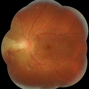
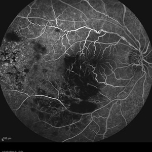
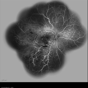
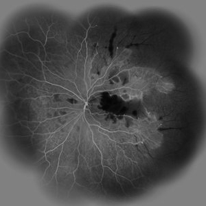 Acute Idiopathic Occlusive Retinal Vasculitis
May 31 2014 by Hamid Ahmadieh, MD
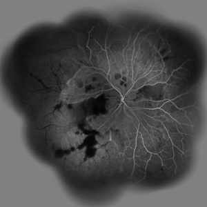
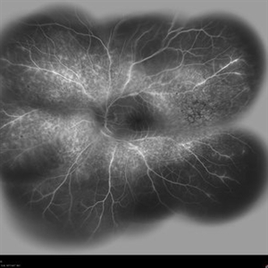
Wide- field fluorescein angiogram of the left eye of a 28-year-old woman with acute drop of vision due to occlusive retinal vasculitis leading to extensive capillary nonperfusion and macular infarction.
Photographer: Naghmeh Nozhat, Negah Eye Center, Tehran
Imaging device: Heidelberg Spectralis
Condition/keywords: capillary nonperfusion, retinal infarction, retinal vasculitis
-
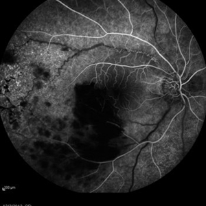
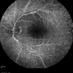
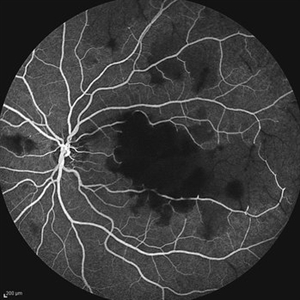 Acute Idiopathic Occlusive Retinal Vasculitis
May 31 2014 by Hamid Ahmadieh, MD
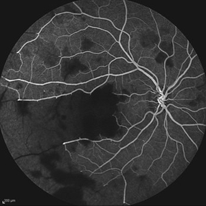
Mid- phase fluorescein angiogram of the left eye of a 28-year-old woman with acute drop of vision due to occlusive retinal vasculitis leading to extensive capillary nonperfusion and macular infarction.
Photographer: Naghmeh Nozhat, Negah Eye Center, Tehran
Imaging device: Heidelberg Spectralis
Condition/keywords: capillary nonperfusion, retinal vasculitis
-
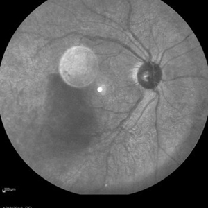
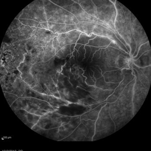
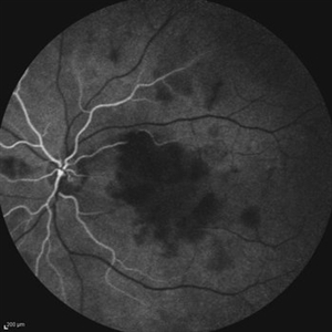 Acute Idiopathic Occlusive Retinal Vasculitis
May 31 2014 by Hamid Ahmadieh, MD
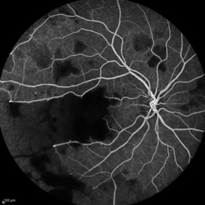
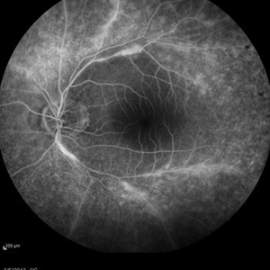
Early phase fluorescein angiogram of the left eye of a 28-year-old woman with acute drop of vision due to occlusive retinal vasculitis leading to extensive capillary nonperfusion and macular infarction.
Photographer: Naghmeh Nozhat, Negah Eye Center, Tehran
Imaging device: Heidelberg Spectralis
Condition/keywords: retinal vasculitis
-
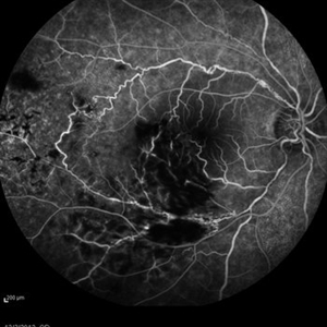
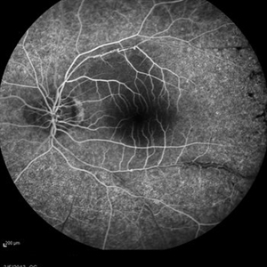
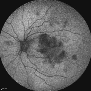 Acute Idiopathic Occlusive Retinal Vasculitis
May 31 2014 by Hamid Ahmadieh, MD
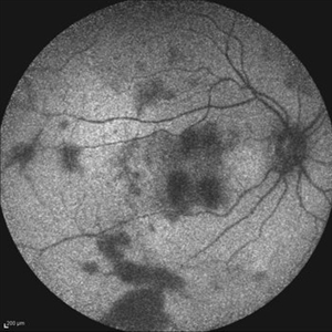
Fundus autofluorescence image of the left eye of a 28-year-old woman with acute drop of vision due to occlusive retinal vasculitis leading to extensive nerve fiber layer infarction and retinal hemorrhages.
Photographer: Naghmeh Nozhat, Negah Eye Center, Tehran
Imaging device: Heidelberg Spectralis
Condition/keywords: fundus autofluorescence (FAF), retinal vasculitis
-
 Acute Idiopathic Occlusive Retinal Vasculitis
May 31 2014 by Hamid Ahmadieh, MD
Wide- field fluorescein angiogram of the right eye of a 28-year-old woman with acute drop of vision due to occlusive retinal vasculitis leading to extensive capillary nonperfusion and macular infarction.
Photographer: Naghmeh Nozhat, Negah Eye Center, Tehran
Imaging device: Heidelberg Spectralis
Condition/keywords: capillary nonperfusion, retinal infarction, retinal vasculitis
-
 Acute Idiopathic Occlusive Retinal Vasculitis
May 31 2014 by Hamid Ahmadieh, MD
Mid phase fluorescein angiogram of the right eye of a 28-year-old woman with acute drop of vision due to occlusive retinal vasculitis leading to extensive capillary nonperfusion and macular infarction.
Photographer: Naghmeh Nozhat, Negah Eye Center, Tehran
Imaging device: Heidelberg Spectralis
Condition/keywords: capillary nonperfusion, retinal vasculitis
-
 Acute Idiopathic Occlusive Retinal Vasculitis
May 31 2014 by Hamid Ahmadieh, MD
Early phase fluorescein angiogram of the right eye of a 28-year-old woman with acute drop of vision due to occlusive retinal vasculitis leading to extensive capillary nonperfusion and macular infarction.
Photographer: Naghmeh Nozhat, Negah Eye Center, Tehran
Imaging device: Heidelberg Spectralis
Condition/keywords: retinal vasculitis
-
 Acute Idiopathic Occlusive Retinal Vasculitis
May 31 2014 by Hamid Ahmadieh, MD
Fundus autofluorescence image of the right eye of a 28-year-old woman with acute drop of vision due to occlusive retinal vasculitis leading to extensive nerve fiber layer infarction and retinal hemorrhages.
Photographer: Naghmeh Nozhat, Negah Eye Center, Tehran
Imaging device: Heidelberg Spectralis
Condition/keywords: fundus autofluorescence (FAF), retinal vasculitis
-
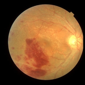
 Acute Toxoplasmosis Neuroretinitis
Mar 15 2017 by Hamid Ahmadieh, MD
Color fundus photograph of the left eye of a 26-year-old man with clinical picture of acute neuroretinitis and serologic evidence of Toxoplasma gondii infection. Disc swelling, necrotizing retinitis with overlying vitreous inflammation, retinal vasculitis and localized exudative retinal detachment are visible.
Photographer: Solmaz Shahmohammad, Negah Eye Center, Tehran,Iran
Condition/keywords: color fundus photograph, exudative retinal detachment, neuroretinitis, retinal vasculitis, toxoplasmosis
-
 Acute Toxoplasmosis Neuroretinitis
Mar 15 2017 by Hamid Ahmadieh, MD
Color fundus photograph of the left eye of a 26-year-old man with clinical picture of acute neuroretinitis and serologic evidence of Toxoplasma gondii infection. Disc swelling, necrotizing retinitis with overlying vitreous inflammation, retinal vasculitis and localized exudative retinal detachment are visible.
Photographer: Solmaz Shahmohammad, Negah Eye Center, Tehran,Iran
Condition/keywords: color fundus photograph, exudative retinal detachment, neuroretinitis, retinal vasculitis
-
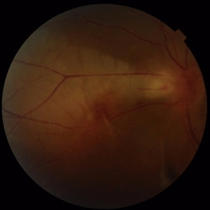
 Behcet's Disease
Mar 13 2013 by Hamid Ahmadieh, MD
Color fundus photograph of the right eye of a 23-year-old man with retinal vasculitis and branch retinal vein occlusion (BRVO) due to Behcet's disease .
Photographer: Solmaz Shahmohammad, Negah Eye Center, Tehran
Imaging device: Heidelberg Spectralis
Condition/keywords: branch retinal vein occlusion (BRVO), retinal vasculitis
-
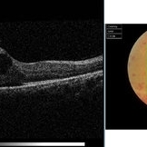 Behcet's Disease
Mar 13 2013 by Hamid Ahmadieh, MD
OCT of the right eye of a 23-year-old man with retinal vasculitis and branch retinal vein occlusion (BRVO) due to Behcet's disease .
Photographer: Solmaz Shahmohammad, Negah Eye Center, Tehran
Imaging device: Topcon OCT
Condition/keywords: branch retinal vein occlusion (BRVO), optical coherence tomography (OCT), retinal vasculitis
-
 Behcet's Disease
Mar 13 2013 by Hamid Ahmadieh, MD
Infrared image of the right eye of a 23-year-old man with retinal vasculitis and branch retinal vein occlusion (BRVO) due to Behcet's disease .
Photographer: Solmaz Shahmohammad, Negah Eye Center, Tehran
Imaging device: Heidelberg Spectralis
Condition/keywords: branch retinal vein occlusion (BRVO), infrared image, retinal vasculitis
-
 Behcet's Disease
Mar 13 2013 by Hamid Ahmadieh, MD
Early phase FA of the right eye of a 23-year-old man with retinal vasculitis and branch retinal vein occlusion (BRVO) due to Behcet's disease .
Photographer: Solmaz Shahmohammad, Negah Eye Center, Tehran
Imaging device: Heidelberg Spectralis
Condition/keywords: branch retinal vein occlusion (BRVO), retinal vasculitis
-
 Behcet's Disease
Mar 13 2013 by Hamid Ahmadieh, MD
Mid phase FA of the right eye of a 23-year-old man with retinal vasculitis and branch retinal vein occlusion (BRVO) due to Behcet's disease .
Photographer: Solmaz Shahmohammad, Negah Eye Center, Tehran
Imaging device: Heidelberg Spectralis
Condition/keywords: branch retinal vein occlusion (BRVO), retinal vasculitis
-
 Behcet's Disease
Mar 13 2013 by Hamid Ahmadieh, MD
Late phase FA of the right eye of a 23-year-old man with retinal vasculitis and branch retinal vein occlusion (BRVO) due to Behcet's disease .
Photographer: Solmaz Shahmohammad, Negah Eye Center, Tehran
Imaging device: Heidelberg Spectralis
Condition/keywords: branch retinal vein occlusion (BRVO), retinal vasculitis
-
 Behcet's Disease
Mar 13 2013 by Hamid Ahmadieh, MD
Wide field FA of the right eye of a 23-year-old man with retinal vasculitis and branch retinal vein occlusion (BRVO) due to Behcet's disease .
Photographer: Solmaz Shahmohammad, Negah Eye Center, Tehran
Imaging device: Heidelberg Spectralis
Condition/keywords: branch retinal vein occlusion (BRVO), retinal vasculitis
-
 Behcet's Disease
Mar 13 2013 by Hamid Ahmadieh, MD
Early phase FA of the left eye of a 23-year-old man with retinal vasculitis due to Behcet's disease .
Photographer: Solmaz Shahmohammad, Negah Eye Center, Tehran
Imaging device: Heidelberg Spectralis
Condition/keywords: retinal vasculitis
-
 Behcet's Disease
Mar 13 2013 by Hamid Ahmadieh, MD
Mid phase FA of the left eye of a 23-year-old man with retinal vasculitis due to Behcet's disease .
Photographer: Solmaz Shahmohammad , Negah Eye Center, Tehran
Imaging device: Heidelberg Spectralis
Condition/keywords: retinal vasculitis
-
 Behcet's Disease
Mar 13 2013 by Hamid Ahmadieh, MD
Late phase FA of the left eye of a 23-year-old man with retinal vasculitis due to Behcet's disease .
Photographer: Solmaz Shahmohammad, Negah Eye Center, Tehran
Imaging device: Heidelberg Spectralis
Condition/keywords: retinal vasculitis
-
 Behcet's Disease
Mar 13 2013 by Hamid Ahmadieh, MD
Wide field FA of the left eye of a 23-year-old man with retinal vasculitis due to Behcet's disease .
Photographer: Solmaz Shahmohammadi , Negah Eye Center, Tehran
Imaging device: Heidelberg Spectralis
Condition/keywords: retinal vasculitis
-
---thumb.jpg/image-square;max$300,300.ImageHandler) Bilateral Retinal Vasculitis
Bilateral Retinal Vasculitis
Oct 29 2013 by Maurice F. Rabb
47 year old white man with bilateral retinal vasculitis.
Condition/keywords: retinal vasculitis
-
---thumb.jpg/image-square;max$300,300.ImageHandler) Bilateral Retinal Vasculitis
Bilateral Retinal Vasculitis
Oct 29 2013 by Maurice F. Rabb
47 year old white man with bilateral retinal vasculitis.
Condition/keywords: retinal vasculitis
-
---thumb.jpg/image-square;max$300,300.ImageHandler) Bilateral Retinal Vasculitis
Bilateral Retinal Vasculitis
Oct 29 2013 by Maurice F. Rabb
47 year old white man with bilateral retinal vasculitis.
Condition/keywords: retinal vasculitis
 Loading…