Search results (100 results)
-
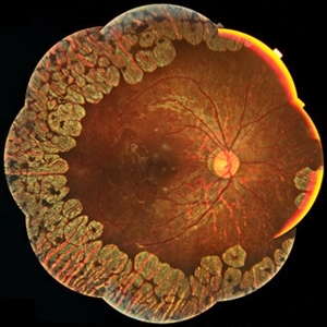
 Asteroid Hyalosis in Retinitis Pigmentosa
Dec 9 2024 by Mauricio Bayram-Suverza, MD
A 54 year-old male patient presented with asteroid hyalosis. Retinal examination revealed the presence of bone spicules, primarily located in the mid-periphery. Genetic testing identified a pathogenic variant in the RHO gene.
Photographer: Mauricio Bayram-Suverza, Casey Eye Institute, OHSU.
Imaging device: Optos California
Condition/keywords: Asteroid hyalosis, retinal dystrophy, Retinitis Pigmentosa, vitreous
-
 Central Areolar Choroidal Dystrophy
Apr 14 2018 by Hamza Ahmed Shawky
Left fundus smartphone photograph of a 35-year-old man with central areolar choroidal dystrophy, BCVA is 6/60
Photographer: Hamza Shawky, Alferdaws eye hospital, Retina unit
Imaging device: smartphone fundus photography
Condition/keywords: central areolar choroidal dystrophy (CACD), hereditary retinal dystrophy, macular dystrophy, retinal dystrophy
-
 Central Areolar Choroidal Dystrophy
Apr 14 2018 by Hamza Ahmed Shawky
Right fundus smartphone photograph of a 35-year-old man with central areolar choroidal dystrophy, BCVA is 6/60
Photographer: Hamza Shawky, Alferdaws eye hospital, Retina unit
Imaging device: smartphone fundus photography
Condition/keywords: central areolar choroidal dystrophy (CACD), hereditary retinal dystrophy, macular dystrophy, retinal dystrophy
-
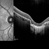
 Central Areolar Choroidal Dystrophy
Apr 14 2018 by Hamza Ahmed Shawky
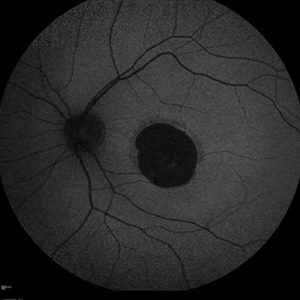
Left fundus OCT of a 35-year-old man with central areolar choroidal dystrophy, BCVA is 6/60
Photographer: Hamza Shawky, Alferdaws eye hospital, Retina unit
Imaging device: Heidelberg Spectralis
Condition/keywords: central areolar choroidal dystrophy (CACD), hereditary retinal dystrophy, macular dystrophy, retinal dystrophy
-
 Central Areolar Choroidal Dystrophy
Apr 14 2018 by Hamza Ahmed Shawky
Right fundus OCT of a 35-year-old man with central areolar choroidal dystrophy, BCVA is 6/60
Photographer: Hamza Shawky, Alferdaws eye hospital, Retina unit
Imaging device: Heidelberg Spectralis
Condition/keywords: central areolar choroidal dystrophy (CACD), hereditary retinal dystrophy, macular dystrophy, retinal dystrophy
-
 Central areolar choroidal dystrophy
Apr 14 2018 by Hamza Ahmed Shawky
Right fundus autofluorescence photograph of a 35-year-old man with central areolar choroidal dystrophy, BCVA is 6/60
Photographer: Hamza Shawky, Alferdaws eye hospital, Retina unit
Imaging device: Heidelberg Spectralis
Condition/keywords: central areolar choroidal dystrophy (CACD), hereditary retinal dystrophy, macular dystrophy, retinal dystrophy
-
 Central Areolar Choroidal Dystrophy
Apr 14 2018 by Hamza Ahmed Shawky
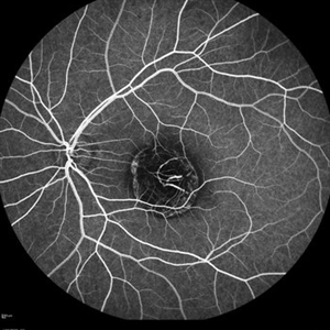
Right fundus late FFA photograph of a 35-year-old man with central areolar choroidal dystrophy, BCVA is 6/60
Photographer: Hamza Shawky, Alferdaws eye hospital, Retina unit
Imaging device: Heidelberg Spectralis
Condition/keywords: central areolar choroidal dystrophy (CACD), hereditary retinal dystrophy, macular dystrophy, retinal dystrophy
-
 Central Areolar Choroidal Dystrophy
Apr 14 2018 by Hamza Ahmed Shawky
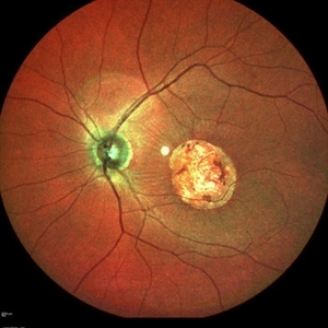
Right fundus color photograph of a 35-year-old man with central areolar choroidal dystrophy, BCVA is 6/60
Photographer: Hamza Shawky, Alferdaws eye hospital, Retina unit
Imaging device: Heidelberg Spectralis
Condition/keywords: central areolar choroidal dystrophy (CACD), hereditary retinal dystrophy, macular dystrophy, retinal dystrophy
-
 Central Areolar Choroidal Dystrophy
Apr 14 2018 by Hamza Ahmed Shawky
Left fundus late FFA photograph of a 35-year-old man with central areolar choroidal dystrophy, BCVA is 6/60
Photographer: Hamza Shawky, Alferdaws eye hospital, Retina unit
Imaging device: Heidelberg Spectralis
Condition/keywords: central areolar choroidal dystrophy (CACD), hereditary retinal dystrophy, macular dystrophy, retinal dystrophy
-
 Central Areolar Choroidal Dystrophy
Apr 14 2018 by Hamza Ahmed Shawky
Left fundus autofluorescence photograph of a 35-year-old man with central areolar choroidal dystrophy, BCVA is 6/60
Photographer: Hamza Shawky, Alferdaws eye hospital, Retina unit
Imaging device: Heidelberg Spectralis
Condition/keywords: central areolar choroidal dystrophy (CACD), hereditary retinal dystrophy, macular dystrophy, retinal dystrophy
-
 Central Areolar Choroidal Dystrophy
Apr 14 2018 by Hamza Ahmed Shawky
Left fundus color photograph of a 35-year-old man with central areolar choroidal dystrophy, BCVA is 6/60.
Photographer: Hamza Shawky, Alferdaws eye hospital, Retina unit
Imaging device: Heidelberg Spectralis
Condition/keywords: central areolar choroidal dystrophy (CACD), hereditary retinal dystrophy, macular dystrophy, retinal dystrophy
-
 Cone-Rod Dystrophy
Jul 20 2023 by Harsh Vardhan Singh, MS
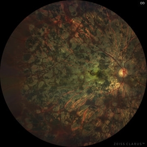
52-year-old male with a advanced stage of cone-rod dystrophy
Photographer: Harsh Vardhan Singh, AIIMS, Guwahati
Imaging device: Zeiss Clarus 700
Condition/keywords: cone dystrophy, Cone-Rod Dystrophy, pigmentary retinal dystrophy, retinal dystrophy
-
 Gyrate Atrophy
Jan 6 2019 by Hashim Ali Khan, OD, FAAO
Montage of Multiple Fundus Photographs from the right eye of a 25-year-old woman with gyrate atrophy.
Photographer: Ahmed Abbass
Imaging device: Topcon TRC-NW8F
Condition/keywords: gyrate atrophy, hereditary retinal dystrophy, retinal dystrophy
-
 Leber´s Congenital Amaurosis
Sep 6 2024 by Mauricio Bayram-Suverza, MD
13-year-old female patient with severe nyctalopia, photophobia, and reduced peripheral vision. CRB1-related Leber´s Congenital Amaurosis. The ultra-widefield pseudocolor image shows attenuated arterioles and diffuse nummular pigmentation with important atrophy.
Photographer: Mauricio Bayram-Suverza, Casey Eye Institute, OHSU.
Imaging device: Optos California
Condition/keywords: genetic testing, Leber's congenital amaurosis, nyctalopia, retinal dystrophy
-
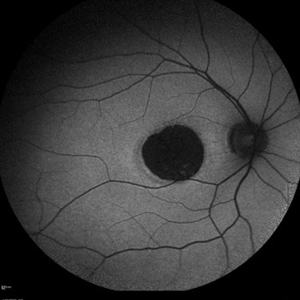
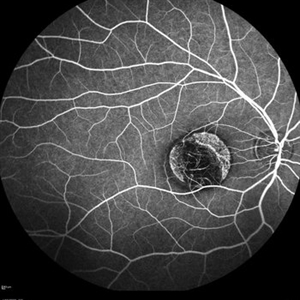
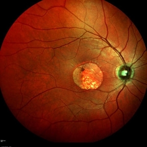
 Pericentral Retinitis Pigmentosa
Sep 6 2024 by Mauricio Bayram-Suverza, MD
A 65-year-old male patient reports experiencing bilateral blind spots that have gradually intensified over time. Genetic testing was unrevealing. The fundus autofluorescence image shows a hypoautofluorescent ring in the posterior pole, especially nasal to the nerve and along arcades.
Photographer: Mauricio Bayram-Suverza, Casey Eye Institute, OHSU.
Imaging device: Optos California
Condition/keywords: fundus autofluorescence (FAF), inherited retinal disease, nyctalopia, retinal dystrophy, retinitis pigmentosa
-
---thumb.jpg/image-square;max$300,300.ImageHandler) Retinal Dystrophy
Retinal Dystrophy
Aug 9 2013 by From the Collections of Thomas M. Aaberg, MD and Thomas M. Aaberg Jr., MD
Pigmented dystrophy.
Condition/keywords: pigmentary retinal dystrophy, retinal dystrophy
-
---thumb.jpg/image-square;max$300,300.ImageHandler) Retinal Dystrophy
Retinal Dystrophy
Aug 9 2013 by From the Collections of Thomas M. Aaberg, MD and Thomas M. Aaberg Jr., MD
Pigmented dystrophy.
Condition/keywords: pigmentary retinal dystrophy, retinal dystrophy
-
---thumb.jpg/image-square;max$300,300.ImageHandler) Retinal Dystrophy
Retinal Dystrophy
Aug 9 2013 by From the Collections of Thomas M. Aaberg, MD and Thomas M. Aaberg Jr., MD
FA of central atrophy with flecks.
Condition/keywords: central choroidal atrophy, total, flecks, retinal dystrophy
-
---thumb.jpg/image-square;max$300,300.ImageHandler) Retinal Dystrophy
Retinal Dystrophy
Aug 9 2013 by From the Collections of Thomas M. Aaberg, MD and Thomas M. Aaberg Jr., MD
Large PED.
Condition/keywords: pigment epithelial detachment (PED), retinal dystrophy
-
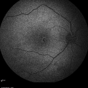 Retinal Dystrophy of 24-Year-Old Male Early FA OD
Nov 25 2015 by Zach Dupureur
Fluorescein angiography of a 24-year-old male. Juvenile retinoschisis on OCT. FA shows outer retinal staining. Could be associated with Goldmann Farve Syndrome.
Condition/keywords: Goldmann-Favre Syndrome, juvenile retinoschisis, retinal dystrophy
-
 Retinal Dystrophy of 24-Year-Old Male Early FA OD
Nov 25 2015 by Zach Dupureur
Fluorescein angiography of a 24-year-old male. Juvenile retinoschisis on OCT. FA shows outer retinal staining. Could be associated with Goldmann Farve Syndrome.
Photographer: Zach Dupureur, OCT-C
Condition/keywords: Goldmann-Favre Syndrome, juvenile retinoschisis, retinal dystrophy
-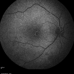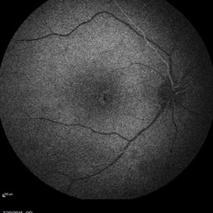
 Retinal Dystrophy of 24-Year-Old Male Early FA OD
Nov 25 2015 by Zach Dupureur
Fluorescein angiography of a 24-year-old male. Juvenile retinoschisis on OCT. FA shows outer retinal staining. Could be associated with Goldmann Farve Syndrome.
Photographer: Zach Dupureur, OCT-C
Condition/keywords: Goldmann-Favre Syndrome, juvenile retinoschisis, retinal dystrophy
 Loading…