Search results (126 results)
-
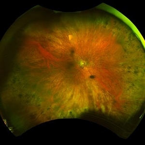
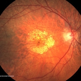 ARMD With Geographic Atrophy, Peripheral Degeneration
Dec 6 2013 by James B. Soque, CRA, OCT-C, COA, FOPS
92-year-old white female with exudative macular degeneration, geographic atrophy, and peripheral retinal degeneration.
Photographer: James Soque, CRA COA, Island Retina, Shirley, New York
Imaging device: Topcon TRC 50DX with OIS 10.6.45
Condition/keywords: geographic atrophy, macular degeneration, retinal degeneration
-
 Bietti's Crystalline Dystrophy
Oct 3 2020 by SHISHIR VERGHESE, MS, FVRS, FAICO (Retina)
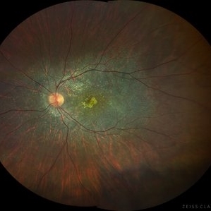
Fundus photo of the left eye of a 21-year-old gentleman with complaints of reduced vision, nyctalopia and visual field reduction. Fundus photograph showing numerous small glistening yellow-white retinal crystalline deposits in the retina.
Photographer: Shishir Verghese, Aravind Eye Hospital, Coimbatore
Condition/keywords: Bietti's crystalline dystrophy, heredomacular degeneration, retinal degeneration
-
 Giant Retinal Tear
Oct 9 2012 by Audina M. Berrocal, MD FASRS
Teenager with high myopia and a GRT
Photographer: Ditte Hess CRA, BPEI
Imaging device: Fundus Camera
Condition/keywords: high myopia, retinal degeneration, retinal tear
-
 Operculated Retinal Hole
Oct 12 2012 by Jeffrey G. Gross, MD, FASRS
Operculated retinal hole with RD.
Condition/keywords: operculated retinal hole, retinal degeneration
-
 Operculated Retinal Hole in Retinal Detachment
Oct 12 2012 by Jeffrey G. Gross, MD, FASRS
Operculated retinal hole in retinal detachment.
Condition/keywords: operculated retinal hole, retinal degeneration
-
---thumb.jpg/image-square;max$300,300.ImageHandler) RD Repair Cartoon
RD Repair Cartoon
Feb 13 2013 by From the Collections of Thomas M. Aaberg, MD and Thomas M. Aaberg Jr., MD
Extrusion needle, endoilluminator.
Condition/keywords: cartoon, enclosed ora bay, extrusion needle, retinal degeneration
-
---thumb.jpg/image-square;max$300,300.ImageHandler) Retinal Degeneration
Retinal Degeneration
Oct 7 2013 by Maurice F. Rabb
Nine year old Afro American girl who was referred for the evaluation of a retinal degeneration. Visual acuity was correctable to 20/60 in each eye. Intraocular pressures where 13 mmHg OD and 12 mmHg OS. There was a trace to 1 + cells in the right vitreous and +1 to +2 cells in the left. Fundus exam showed evidence for diffuse RPE hypopigmentation. The changes were consistent with a diffuse pigmentary degeneration of the retina in each eye. The patient's retinal degeneration is associated with a systemic syndrome.
Condition/keywords: retinal degeneration
-
 Retinal Detachment
Nov 9 2012 by Norman Byer
This is a retinal detachment in a 55-year-old man. The vertical convex line on the right side probably represents the posterior border of the vitreous base. Note the small tractional tear with the base of its flap attached at this line. This demonstrates how the vitreous base presents an effective barrier to further extension of the retinal tear. Note also how the flap breaks the continuity of the yellow line.
Condition/keywords: retinal degeneration, retinal flap, tractional retinal tear, vitreous base
-
 Retinal Detachment with Macula Partially Detached
Oct 12 2012 by Jeffrey G. Gross, MD, FASRS
RD with macula partially detached.
Condition/keywords: macula, retinal degeneration
-
 Rhegmatogenous Retinal Detachment
Oct 5 2012 by Ronald C. Gentile, MD
Rhegmatogenous retinal detachment involving the macula with characteristic outer hydration lines and associated peripheral retinal tear (top left of image). The choroidal vasculature can be seen through the break in the retina.
Photographer: The New York Eye & Ear Infirmary Department of Medical Imaging
Condition/keywords: retinal degeneration
-
 Rhegmatogenous Retinal Detachment
Oct 5 2012 by Ronald C. Gentile, MD
Rhegmatogenous retinal detachment (right eye, temporal retina) involving the macula with characteristic outer hydration lines.
Photographer: The New York Eye & Ear Infirmary Department of Medical Imaging
Condition/keywords: retinal degeneration
-
 Serpiginous retinal degeneration
Jul 19 2019 by JEFFERSON R SOUSA, Tecg.º (Biomedical Systems Technology)
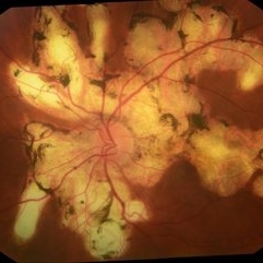
54-year-old female patient, visual acuity 20/100 both eyes. In the evaluation, she presented choroidal atrophy, starting from the optic nerve towards the periphery of the retina. Serpiginous retinal degeneration
Photographer: JEFFERSON R SOUSA - Study Center and Ophthalmological Research Dr. Andre M V Gomes, Institute Dr. Suel Abujamra São Paulo-Brazil
Imaging device: Topcon TRC-50 DX, Imaginet 4.0, angle de 35 graus. Flash 36 w-s
Condition/keywords: retinal degeneration, serpiginous
-
 Serpiginous Retinal Degeneration
Jul 19 2019 by JEFFERSON R SOUSA, Tecg.º (Biomedical Systems Technology)
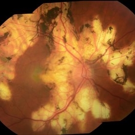
54-year-old female patient, visual acuity 20/100 both eyes. In the evaluation, she presented choroidal atrophy, starting from the optic nerve towards the periphery of the retina. Serpiginous retinal degeneration
Photographer: JEFFERSON R SOUSA - Study Center and Ophthalmological Research Dr. Andre M V Gomes, Institute Dr. Suel Abujamra São Paulo-Brazil
Imaging device: Topcon TRC-50 DX, Imaginet 4.0, angle de 35 graus. Flash 36 w-s
Condition/keywords: retinal degeneration, serpiginous
-
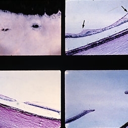 Slide 8-9
Mar 4 2019 by Lancaster Course in Ophthalmology
Lattice degeneration of retina with retinal holes. Microscopic appearance at three levels (upper, right, and lower) shows discontinuity of the internal limiting membrane, an overlying pocket of fluid vitreous, atrophy of inner retinal layers, hole formation, and vitreoretinal adhesion (arrows) at margin of lesion. (E.P. No. 30544)
Condition/keywords: atrophy, retinal degeneration, retinal hole, vitreoretinal adhesion
-
 Unknown
Jul 23 2013 by Howard Schatz, MD
RD 20, angioma.
Condition/keywords: angioma, retinal degeneration, unknown
-
 Alagille Syndrome UWF Autofluorescence
Dec 4 2023 by Isaac Ezon, MD
43 yo Female with known Alagille Syndrome, referred for peripheral retinal changes. Subjective nyctalopia but no other symtpoms. Alagille Syndrome UWF Autofluorescence.
Photographer: Tara Murray
Imaging device: Optos
Condition/keywords: hereditary choroidal dystrophy, hereditary retinal degeneration
-
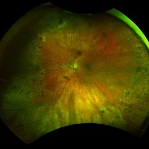 Alagille Syndrome UWF Color
Dec 4 2023 by Isaac Ezon, MD
43 yo Female with known Alagille Syndrome, referred for peripheral retinal changes. Subjective nyctalopia but no other symtpoms. Alagille Syndrome UWF Color.
Photographer: Tara Murray
Imaging device: Optos
Condition/keywords: hereditary choroidal dystrophy, hereditary retinal degeneration
-
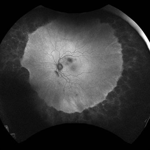
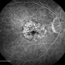 ARMD With Geographic Atrophy , Peripheral Degeneration, FA
Dec 6 2013 by James B. Soque, CRA, OCT-C, COA, FOPS
FA right eye early phase, of a 92-year-old white female with exudative macular degeneration, geographic atrophy, and peripheral retinal degeneration.
Photographer: James Soque CRA COA, Island Retina, Shirley, New York
Imaging device: Topcon TRC 50DX with OIS 10.6.45
Condition/keywords: geographic atrophy
-
 ARMD With Geographic Atrophy, Peripheral Degeneration
Dec 6 2013 by James B. Soque, CRA, OCT-C, COA, FOPS
92-year-old white female with exudative macular degeneration, geographic atrophy, and peripheral retinal degeneration.
Photographer: James Soque, CRA COA, Island Retina, Shirley, New York
Imaging device: Topcon TRC 50DX with OIS 10.6.45
Condition/keywords: fundus photograph, geographic atrophy
-
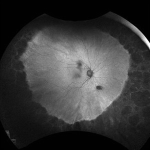
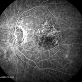 ARMD With Geographic Atrophy, Peripheral Degeneration, FA
Dec 6 2013 by James B. Soque, CRA, OCT-C, COA, FOPS
FA left eye Early Phase, of a 92-year-old white female with exudative macular degeneration, geographic atrophy, and peripheral retinal degeneration.
Photographer: James Soque CRA COA, Island Retina, Shirley, New York
Imaging device: Topcon TRC 50DX with OIS 10.6.45
Condition/keywords: geographic atrophy
 Loading…