Search results (37 results)
-
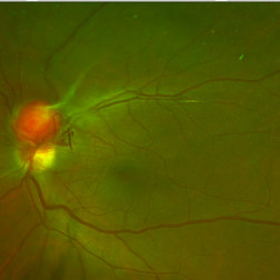
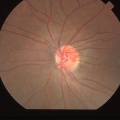
 Retinal Capillary Hemangioma
May 31 2017 by S. Natarajan, MD, FASRS, FRCS (GLASGOW) , FICO, D.Sc, FELA
Fundus photograph of an 21-year-old male with double angioma before undergoing laser photo ablation.
Photographer: Ms. Ashwini Borde
Imaging device: Carl Zeiss 450 Plus IR
Condition/keywords: angioma, disc
-
 Retinal Capillary Hemangioma
Mar 21 2013 by Yusuke Oshima, MD, PhD
A peripheral retinal capillary hemangioma.
Photographer: Yusuke Takada, Osaka University Graduate School of Medicine
-
---thumb.jpg/image-square;max$300,300.ImageHandler) Retinal Capillary Hemangioma
Retinal Capillary Hemangioma
May 3 2013 by Jerald A. Bovino, MD
Fundus photo of a retinal capillary hemangioma after treatment.
-
---thumb.jpg/image-square;max$300,300.ImageHandler) Retinal Capillary Hemangioma
Retinal Capillary Hemangioma
May 3 2013 by Jerald A. Bovino, MD
Fundus photo of a retinal capillary hemangioma after treatment of the feeder vessel.
Condition/keywords: feeder vessel
-
 Retinal Capillary Hemangioma
May 3 2013 by Jerald A. Bovino, MD
Fundus photo of a retinal capillary hemangioma after treatment of the feeder vessel.
Condition/keywords: feeder vessel
-
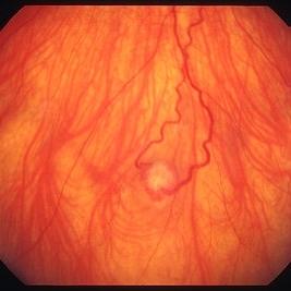
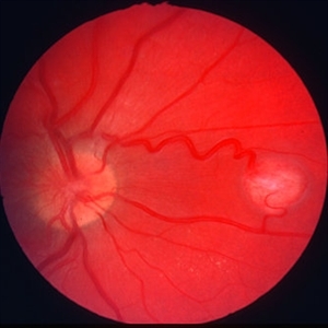 Retinal capillary hemangioma
Jan 11 2013 by Alex P. Hunyor, MD
Retinal capillary haemangioma nasal to optic disc, right eye.
Condition/keywords: Von Hippel-Lindau
-
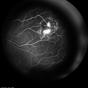 Retinal Capillary Hemangioma
Feb 18 2016 by Hashim Ali Khan, OD, FAAO
FA of 23-year-old-woman with RCH as part of spectrum of Von Hippel- Lindau
Imaging device: Heidelberg Spectralis
Condition/keywords: Von Hippel-Lindau
-
 Retinal Capillary Hemangioma
Feb 10 2016 by Claudia G Hooten, MD
Fundus photo of 47-year-old male with PVR retinal detachment 2 months post cryotherapy of RCH.
Photographer: Mark D. Clark
-
 Retinal Capillary Hemangioma
Nov 9 2016 by Courtney Crawford, MD, FACS
Fundus photograph of a 30-year-old woman with a retinal capillary hemangioma and known Von Hippel-Lindau.
Condition/keywords: Von Hippel-Lindau
-
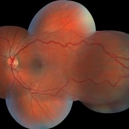
 Retinal Capillary Hemangioma
Dec 12 2019 by David L Kilpatrick, MD
This is a wide-field color fundus photo showing two distinct retinal capillary hemangiomas. A visually significant epiretinal membrane is also present. Work up with gene testing was negative for VHL. The plan is to proceed with PDT of the two separate lesions (half fluence for the peripapillary lesion), followed by cryotherapy / photocoagulation.
Photographer: Retina Consultants of Alabama
Imaging device: Optos
-
 Retinal Capillary Hemangioma
Dec 12 2019 by David L Kilpatrick, MD
This is a wide-field color fundus photo showing two distinct retinal capillary hemangiomas. A visually significant epiretinal membrane is also present. Work up with gene testing was negative for VHL. The plan is to proceed with PDT of the two separate lesions (half fluence for the peripapillary lesion), followed by cryotherapy / photocoagulation.
-
 Retinal Capillary Hemangioma
Dec 12 2019 by David L Kilpatrick, MD
OCT showing two distinct retinal capillary hemangiomas. A visually significant epiretinal membrane is also present. Work up with gene testing was negative for VHL. The plan is to proceed with PDT of the two separate lesions (half fluence for the peripapillary lesion), followed by cryotherapy / photocoagulation.
-
 Retinal Capillary Hemangioma
Aug 2 2017 by Nathalia Roberti
Wide fundus photo of a 21-year-old man with capillary hemagioma in the left eye. His best-corrected visual acuity was 20/20.
Imaging device: Visucam
Condition/keywords: hemangioma
-
 Retinal Capillary Hemangioma
Sep 9 2021 by Jesus Lozano, MD
60 year-old woman with a Peripheral RCH treated with laser photocoagulation.
Photographer: Yair Bet Yosef, Hadassah Medical Center. Israel
Imaging device: Optos Silverstone
Condition/keywords: abnormal retinal vessel, anomalous vessels, dilated tortuous vessels, hemangioma, retina
-
 Retinal Capillary Hemangioma
Jan 20 2021 by Jamin S. Brown, MD
Retinal capillary hemangioma, OD.
Photographer: Stefanie Palmer CRA, Retina Vitreous Surgeons of CNY
Condition/keywords: fluorescein angiogram (FA)
-
 Retinal capillary hemangioma 2
Jan 11 2013 by Alex P. Hunyor, MD
Retinal capillary haemangioma, right inferior periphery, in a 20-year-old female with von Hippel-Lindau disease.
Condition/keywords: hemangioma, Von Hippel-Lindau
-
---thumb.jpg/image-square;max$300,300.ImageHandler) Retinal capillary hemangioma 4 image 1
Retinal capillary hemangioma 4 image 1
Jan 11 2013 by Alex P. Hunyor, MD
Retinal capillary haemangioma, left eye, in a young female with von Hippel-Lindau disease. Color image 1 showing extensive lipid deposition in the macula.
Condition/keywords: Von Hippel-Lindau
-
---thumb.jpg/image-square;max$300,300.ImageHandler) Retinal capillary hemangioma 4 image 2
Retinal capillary hemangioma 4 image 2
Jan 11 2013 by Alex P. Hunyor, MD
Retinal capillary haemangioma, left eye, in a young female with von Hippel-Lindau disease. Color image 2 showing the haemangioma with surrounding exudative detachment and lipid exudate.
Condition/keywords: Von Hippel-Lindau
-
---thumb.JPG/image-square;max$300,300.ImageHandler) Retinal Capillary Hemangioma Due to Von Hippel-Lindau Syndrome
Retinal Capillary Hemangioma Due to Von Hippel-Lindau Syndrome
Jul 11 2013 by Jason S. Calhoun
A 47-year-old female in for a second opinion for retinal capillary hemangioma in the left eye. VA was HM (hand motion) in the left eye. She has had multiple procedures done for the left eye including, retinal detachment surgery, YAG Laser PI, YAG laser capsulotomy, and cataract surgery.
Photographer: Jason S. Calhoun, Department of Ophthalmology, Mayo Clinic Jacksonville, Florida
-
 Retinal Capillary Hemangioma in Von Hippel-Lindau
Feb 7 2016 by David B Lazar, MD
26-year-old woman with Von Hippel-Lindau being followed for multiple pancreatic cysts, renal cysts and renal cell carcinomas.
Photographer: Timothy Tivnan, Lahey Clinic Foundation, Burlington, MA
Imaging device: Optos Wide-field Imaging
Condition/keywords: Von Hippel-Lindau
-
 Retinal capillary hemangiomas 3
Jan 11 2013 by Alex P. Hunyor, MD
Retinal capillary haemangiomas, left superior periphery, in a 20 year old female with von Hippel-Lindau disease.
Condition/keywords: hemangioma, Von Hippel-Lindau
-
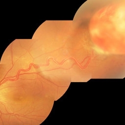
 Atypical Arterial Beading Secondary to Retinal Capillary Hemangioma
Jan 28 2020 by Sophia El Hamichi, MD
Image 1: Fundus picture montage depicting RCH with feeder and drainer vessels. Note the unusual beaded appearance of the arterioles. Image 2: 2A: Arterial phase of fluorescein angiography shows early filling of the arteriole. 2B: Arterio-venous phase highlighting the sausage appearance of the arterioles beading.
Condition/keywords: arterial beading, fluorescein angiogram (FA), retinal capillary hemangioblastoma
-
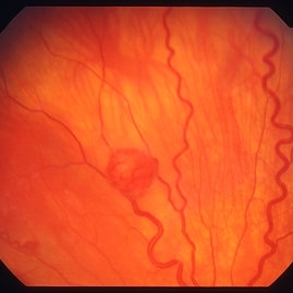
 Capillary Hemangioma
Mar 27 2019 by Gary R. Cook, MD, FACS
White male with a retinal capillary hemangioma OD secondary to neurofibromatosis.
Condition/keywords: neurofibromatosis, retinal hemangioma
-
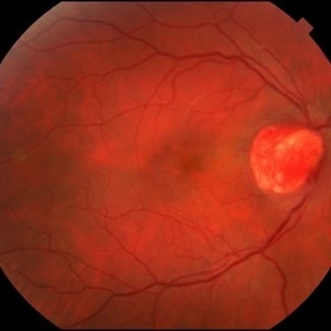
 Capillary Hemangioma
Mar 27 2019 by Gary R. Cook, MD, FACS
28-year-old asymptomatic white female with a retinal capillary hemangioma of the optic disc OS; V.A.= 20/20-1.
Imaging device: Topcon VT-50
Condition/keywords: optic disc
 Loading…