Search results (51 results)
-
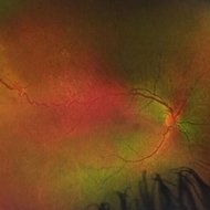
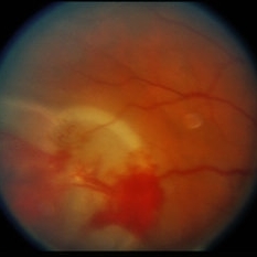
 Atypical Arterial Beading Secondary to Retinal Capillary Hemangioma
Jan 28 2020 by Sophia El Hamichi, MD
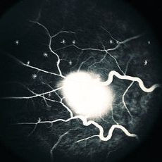
Image 1: Fundus picture montage depicting RCH with feeder and drainer vessels. Note the unusual beaded appearance of the arterioles. Image 2: 2A: Arterial phase of fluorescein angiography shows early filling of the arteriole. 2B: Arterio-venous phase highlighting the sausage appearance of the arterioles beading.
Condition/keywords: arterial beading, fluorescein angiogram (FA), retinal capillary hemangioblastoma, retinal capillary hemangioma
-
 Isolated Retinal Capillary Hemangioblastoma - Early phase IVFA
Mar 11 2022 by Bryon R McKay, MD, PhD, FRCSC, DRCPSC - Retina
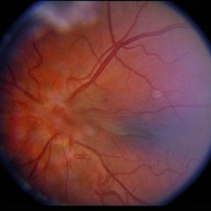
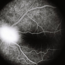
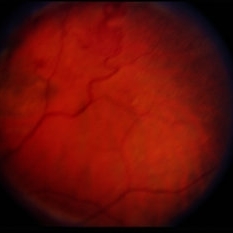
Optos widefield fundus photograph and IVFA of a 23-year-old female with asymptomatic isolated retinal capillary hemangioblastoma without exudation. IVFA demonstrates some mild late leakage. The tumor measures 1.5mm and was effectively ablated with laser photocoagulation.
Imaging device: Optos
Condition/keywords: retinal capillary hemangioblastoma
-
 Isolated Retinal Capillary Hemangioblastoma - A-V phase IVFA
Mar 11 2022 by Bryon R McKay, MD, PhD, FRCSC, DRCPSC - Retina
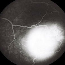
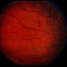
Optos widefield fundus photograph and IVFA of a 23-year-old female with asymptomatic isolated retinal capillary hemangioblastoma without exudation. IVFA demonstrates some mild late leakage. The tumor measures 1.5mm and was effectively ablated with laser photocoagulation.
Imaging device: Optos
Condition/keywords: retinal capillary hemangioblastoma
-
 Isolated Retinal Capillary Hemangioblastoma - Late phase IVFA
Mar 11 2022 by Bryon R McKay, MD, PhD, FRCSC, DRCPSC - Retina
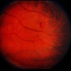
Optos widefield fundus photograph and IVFA of a 23-year-old female with asymptomatic isolated retinal capillary hemangioblastoma without exudation. IVFA demonstrates some mild late leakage. The tumor measures 1.5mm and was effectively ablated with laser photocoagulation.
Imaging device: Optos
Condition/keywords: retinal capillary hemangioblastoma
-
 RCH-OD
Jul 28 2023 by Mohammadkarim Johari
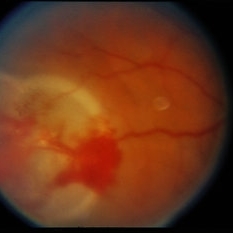
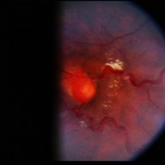
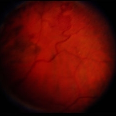
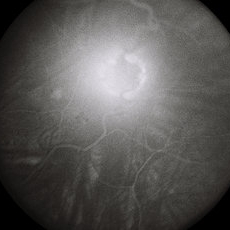
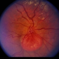
15 year old girl with bilateral vision loss, in right eye a nodular, orange-colored lesions that grow in the outer layers of the retina is seen in supra-temporal quadrant of retina. Retinal capillary hemangioma is a benign retinal hamartoma that may be associated with von Hippel-Lindau (VHL) disease
Photographer: Mohammadkarim Johari, Shiraz university of medical science
Condition/keywords: retinal capillary hemangioblastoma, retinal capillary hemangioma
-
 Retinal Capillary Hemangioblastoma
Jan 12 2022 by VISHAL R RAVAL, MBBS, DNB, FMRF, FASRS
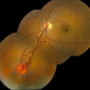
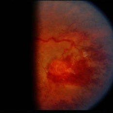
Left eye color fundus photograph of an 42-year-old woman with a large retinal capillary hemangioblastoma tumor with dilated feeder artery and draining vein in the temporal quadrant. There was also presence of tractional retinal detachment with massive intraretinal exudation involving the macula.
Photographer: Bojja Ramesh, LV Prasad Eye Institute, Hyderabad, India
Imaging device: Zeiss Clarus 700
Condition/keywords: intraretinal exudation, retina capillary hemangioblastoma, tractional retinal detachment
-
 Retinal Capillary Hemangioblastoma
May 3 2018 by Nichole Lewis
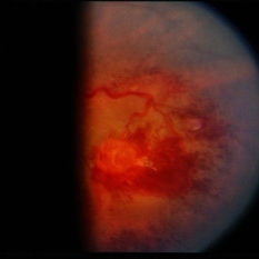
Retinal capillary hemangioblastoma with lipid surrounding.
Photographer: Nichole Lewis
Condition/keywords: lipid exudation, retinal capillary hemangioblastoma
-
 Retinal Capillary Hemangioblastoma
Nov 18 2019 by Megan M Nichols, MD
Fundus photo of a 42-year-old woman who presented without ocular symptoms and was found to have an incidental retinal capillary hemangioblastoma. There was no known history of Von Hippel-Lindau disease.
Photographer: Jonathan Rosen - Beth Israel Lahey Health
Imaging device: TopCon 50DX
Condition/keywords: retinal capillary hemangioblastoma, Von Hippel-Lindau
-
 Retinal Capillary Hemangioblastoma
Feb 20 2015 by H. Michael Lambert, MD
Retinal capillary hemangioblastoma associated with Von Hippel-Lindau Disease. No history. Whitening around lesion possibly cryothermy or laser.
Condition/keywords: retinal capillary hemangioblastoma
-
 Retinal Capillary Hemangioblastoma
Feb 20 2015 by H. Michael Lambert, MD
Retinal capillary hemangioblastoma associated with Von Hippel-Lindau Disease. No history. Dilated tortuous vessel and hemorrhage is shown.
Condition/keywords: retinal capillary hemangioblastoma
-
 Retinal Capillary Hemangioblastoma
Feb 20 2015 by H. Michael Lambert, MD
Retinal capillary hemangioblastoma associated with Von Hippel-Lindau Disease. No history. Whitening around lesion possibly cryothermy or laser.
Condition/keywords: retinal capillary hemangioma
-
 Retinal Capillary Hemangioblastoma
Feb 20 2015 by H. Michael Lambert, MD
Retinal capillary hemangioblastoma associated with Von Hippel-Lindau Disease. No history. Dilated tortuous vessel and hemorrhage is shown.
Condition/keywords: retinal capillary hemangioblastoma, Von Hippel-Lindau
-
 Retinal Capillary Hemangioblastoma
Feb 20 2015 by H. Michael Lambert, MD
Retinal capillary hemangioblastoma associated with Von Hippel-Lindau Disease. No history. Dilated, tortuous afferent and efferent vessels are shown.
Condition/keywords: retinal capillary hemangioblastoma
-
 Retinal Capillary Hemangioblastoma
Feb 20 2015 by H. Michael Lambert, MD
Fluorescein angiogram of a retinal capillary hemangioblastoma associated with Von Hippel-Lindau Disease. No history. Dilated, tortuous afferent and efferent vessels are shown. Fluorescein leakage is beginning in the rather early frame.
Condition/keywords: retinal capillary hemangioblastoma
-
 Retinal Capillary Hemangioblastoma
Feb 20 2015 by H. Michael Lambert, MD
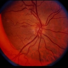
Optic nerve in this eye with fibrosis over the nerve and posterior pole.
Condition/keywords: retinal capillary hemangioblastoma
-
 Retinal Capillary Hemangioblastoma
Feb 20 2015 by H. Michael Lambert, MD
Fluorescein angiogram of a retinal capillary hemangioblastoma No history. Fluorescein leakage is abundant in this later frame.
Condition/keywords: retinal capillary hemangioblastoma
-
 Retinal Capillary Hemangioblastoma
Feb 20 2015 by H. Michael Lambert, MD
Fluorescein angiogram of a retinal capillary hemangioblastoma No history. Fluorescein leakage is substantial in this mid-phase frame.
Condition/keywords: retinal capillary hemangioblastoma
-
 Retinal Capillary Hemangioblastoma
Feb 20 2015 by H. Michael Lambert, MD
Retinal capillary hemangioblastoma associated with Von Hippel-Lindau Disease. No history. Dilated, tortuous afferent and efferent vessels are shown.
Condition/keywords: retinal capillary hemangioblastoma
-
 Retinal Capillary Hemangioblastoma
Feb 20 2015 by H. Michael Lambert, MD
Retinal capillary hemangioblastoma associated with Von Hippel-Lindau Disease. No history. Dilated, tortuous afferent and efferent vessels are shown.
Condition/keywords: retinal capillary hemangioblastoma
-
 Retinal Capillary Hemangioblastoma
Feb 20 2015 by H. Michael Lambert, MD
Small retinal capillary hemangioblastoma associated with Von Hippel-Lindau Disease. No history.
Condition/keywords: retinal capillary hemangioblastoma
-
 Retinal Capillary Hemangioblastoma
Feb 20 2015 by H. Michael Lambert, MD
Small retinal capillary hemangioblastoma associated with Von Hippel-Lindau Disease. No history.
Condition/keywords: retinal capillary hemangioblastoma
-
 Retinal Capillary Hemangioblastoma
Feb 20 2015 by H. Michael Lambert, MD
Fluorescein angiogram of a retinal capillary hemangioblastoma associated with Von Hippel-Lindau Disease. No history. Fluorescein leakage is vague and lesion looks somewhat indolent.
Condition/keywords: retinal capillary hemangioblastoma
-
 Retinal Capillary Hemangioblastoma
Feb 20 2015 by H. Michael Lambert, MD
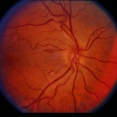
47-year-old with 20/20 OU. Fellow right eye of patient with retinal capillary hemangioblastoma associated the optic nerve in the left eye, in a patient with Von Hippel-Lindau Disease. Small nerve fiber layer hemorrhage is present.
Condition/keywords: retinal capillary hemangioblastoma
-
 Retinal Capillary Hemangioblastoma
Feb 20 2015 by H. Michael Lambert, MD
47-year-old with 20/20 OU. Fellow right eye of patient with retinal capillary hemangioblastoma associated the optic nerve in the left eye, in a patient with Von Hippel-Lindau Disease. Small nerve fiber layer hemorrhage is present.
Condition/keywords: retinal capillary hemangioblastoma
 Loading…