Search results (38 results)
-
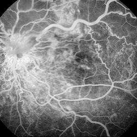
 Angiomatous Retinae
Mar 4 2013 by Judy E. Kim, MD, FARVO, FASRS
Fluorescein angiogram of the lesion in inferior retina.
Condition/keywords: retinal angiomatous proliferation (RAP)
-
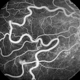
 Choroidal Neovascularization with Retinal Angiomatous Proliferation
Aug 24 2012 by John S. King, MD
Before and 1 week post Avastin; PED, SRF, ME.
Photographer: Kristin Konecki, OcuSight Eye Care Center, Rochester, NY
Condition/keywords: Avastin, retinal angiomatous proliferation (RAP)
-
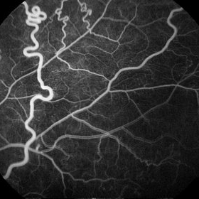
 Choroidal Neovascularization with Retinal Angiomatous Proliferation
Aug 24 2012 by John S. King, MD
16 sec
Photographer: Kristin Konecki, OcuSight Eye Care Center, Rochester, NY
Condition/keywords: retinal angiomatous proliferation (RAP)
-
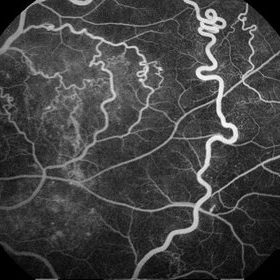
 Choroidal Neovascularization with Retinal Angiomatous Proliferation
Aug 24 2012 by John S. King, MD
1.02 min
Photographer: Kristin Konecki, OcuSight Eye Care Center, Rochester, NY
Condition/keywords: retinal angiomatous proliferation (RAP)
-
 Choroidal Neovascularization with Retinal Angiomatous Proliferation
Aug 24 2012 by John S. King, MD
3.35 min
Photographer: Kristin Konecki, OcuSight Eye Care Center, Rochester, NY
Condition/keywords: retinal angiomatous proliferation (RAP)
-
 Choroidal Neovascularization with Retinal Angiomatous Proliferation
Aug 24 2012 by John S. King, MD
IRH in area of drusen both juxtafoveal and extrafoveal. Photo and FAs are initial presentation
Photographer: Kristin Konecki, OcuSight Eye Care Center, Rochester, NY
Condition/keywords: retinal angiomatous proliferation (RAP)
-
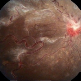
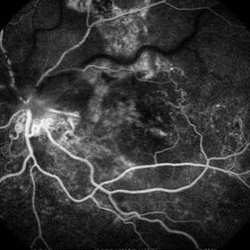
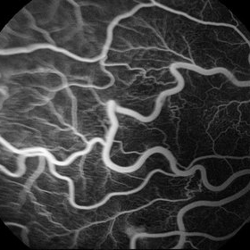
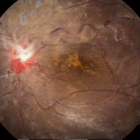 Racemose Hemangioma and Retinal Vein Occlusion
Mar 5 2013 by Eduardo Torres-Porras, MD
A 13-year-old woman had a history of decreased vision for 2 years. Visual acuity was 20/400.
Photographer: Camelia Rosales Lara
Condition/keywords: retinal angiomatous proliferation (RAP)
-
 Racemose Hemangioma and Retinal Vein Occlusion
Mar 5 2013 by Eduardo Torres-Porras, MD
A 13-year-old woman had a history of decreased vision for 2 years. Visual acuity was 20/400.
Photographer: Camelia Rosales Lara
Condition/keywords: retinal angiomatous proliferation (RAP)
-
 Racemose Hemangioma and Retinal Vein Occlusion
Mar 5 2013 by Eduardo Torres-Porras, MD
A 13-year-old woman had a history of decreased vision for 2 years. Visual acuity was 20/400.
Photographer: Camelia Rosales Lara
Condition/keywords: retinal angiomatous proliferation (RAP)
-
 Racemose Hemangioma and Retinal Vein Occlusion
Mar 5 2013 by Eduardo Torres-Porras, MD
A 13-year-old woman had a history of decreased vision for 2 years. Visual acuity was 20/400.
Photographer: Camelia Rosales Lara
Condition/keywords: retinal angiomatous proliferation (RAP)
-
 Racemose Hemangioma and Retinal Vein Occlusion
Mar 5 2013 by Eduardo Torres-Porras, MD
A 13-year-old woman had a history of decreased vision for 2 years. Visual acuity was 20/400.
Photographer: Camelia Rosales Lara
Condition/keywords: retinal angiomatous proliferation (RAP)
-
 Racemose Hemangioma and Retinal Vein Occlusion
Mar 5 2013 by Eduardo Torres-Porras, MD
A 13-year-old woman had a history of decreased vision for 2 years. Visual acuity was 20/400.
Photographer: Camelia Rosales Lara
Condition/keywords: retinal angiomatous proliferation (RAP)
-
 Racemose Hemangioma and Retinal Vein Occlusion
Mar 5 2013 by Eduardo Torres-Porras, MD
A 13-year-old woman had a history of decreased vision for 2 years. Visual acuity was 20/400.
Photographer: Camelia Rosales Lara
Condition/keywords: retinal angiomatous proliferation (RAP)
-
 Racemose Hemangioma and Retinal Vein Occlusion
Mar 5 2013 by Eduardo Torres-Porras, MD
A 13-year-old woman had a history of decreased vision for 2 years. Visual acuity was 20/400.
Photographer: Camelia Rosales Lara
Condition/keywords: retinal angiomatous proliferation (RAP)
-
 Racemose Hemangioma and Retinal Vein Occlusion
Mar 5 2013 by Eduardo Torres-Porras, MD
A 13-year-old woman had a history of decreased vision for 2 years. Visual acuity was 20/400.
Photographer: Camelia Rosales Lara
Condition/keywords: retinal angiomatous proliferation (RAP)
-
 Racemose Hemangioma and Retinal Vein Occlusion
Mar 5 2013 by Eduardo Torres-Porras, MD
A 13-year-old woman had a history of decreased vision for 2 years. Visual acuity was 20/400.
Photographer: Camelia Rosales Lara
Condition/keywords: retinal angiomatous proliferation (RAP)
-
 RAP lesions
Sep 29 2014 by Thomas A. Ciulla, MD, MBA, FASRS
OCT of an 81-year-old man revealing several RAP lesions superior to fovea.
Photographer: Stuart Alfred CRA
Condition/keywords: choroidal neovascular membrane (CNVM), neovascular age-related macular degeneration (AMD), retinal angiomatous proliferation (RAP), wet age-related macular degeneration (wet AMD)
-
 RAP lesions
Sep 29 2014 by Thomas A. Ciulla, MD, MBA, FASRS
Late frame fluorescein angiogram of an 81-year-old man revealing several RAP lesions superior to fovea.
Photographer: Stuart Alfred CRA
Condition/keywords: choroidal neovascular membrane (CNVM), neovascular age-related macular degeneration (AMD), retinal angiomatous proliferation (RAP), wet age-related macular degeneration (wet AMD)
-
 RAP lesions
Sep 29 2014 by Thomas A. Ciulla, MD, MBA, FASRS
Fluorescein angiogram of an 81-year-old man revealing several RAP lesions superior to fovea.
Photographer: Stuart Alfred CRA
Condition/keywords: choroidal neovascular membrane (CNVM), neovascular age-related macular degeneration (AMD), retinal angiomatous proliferation (RAP), wet age-related macular degeneration (wet AMD)
-
 RAP Lesions
Sep 29 2014 by Thomas A. Ciulla, MD, MBA, FASRS
Fluorescein angiogram of an 81-year-old man revealing several RAP lesions superior to fovea.
Photographer: Stuart Alfred CRA
Condition/keywords: choroidal neovascular membrane (CNVM), neovascular age-related macular degeneration (AMD), retinal angiomatous proliferation (RAP), wet age-related macular degeneration (wet AMD)
-
 RAP lesions
Sep 29 2014 by Thomas A. Ciulla, MD, MBA, FASRS
Fluorescein angiogram of an 81-year-old man revealing several RAP lesions superior to fovea.
Photographer: Stuart Alfred CRA
Condition/keywords: choroidal neovascular membrane (CNVM), neovascular age-related macular degeneration (AMD), retinal angiomatous proliferation (RAP), wet age-related macular degeneration (wet AMD)
-
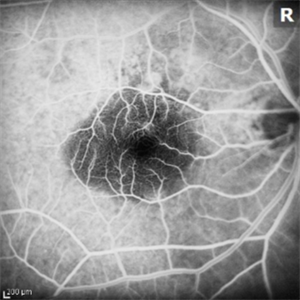
 Retinal Angiomatous Proliferation
Sep 10 2018 by Gabriela Lopezcarasa Hernandez, MD
75-year-old patient with decrease in visual acuity right eye with metamorphopsia, in the FA and ICG we can see a RAP lesion.
Photographer: Azucena Rios
Imaging device: Heidelberg Spectralis
Condition/keywords: FA mid phase, indocyanine green (ICG) angiography, RAP lesion, retinal angiomatous proliferation (RAP)
-
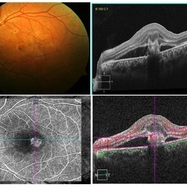
 Retinal Angiomatous Proliferation
Oct 11 2012 by Gabriela Lopezcarasa Hernandez, MD
70-year-old male with diagnostic of RAP by OCT and ICG fluorescein angiography
Photographer: Azucena Rios, Macula Retina Consultores Mexico
Imaging device: Heidelberg Spectralis
Condition/keywords: retinal angiomatous proliferation (RAP)
-
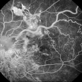
 Retinal Angiomatous Proliferation
May 7 2021 by Dhaivat Shah
Retinal angiomatous proliferation (RAP) is a distinct variant of neovascular age-related degeneration (AMD) that usually initiates at the retina and progresses posteriorly into sub retinal space. In most recent study, it was suggested that angiogenesis may begin in the retina, choroid, or both, and introduced a new name for the process: Type 3 neovascularization. The frequency of RAP has been studied in many studies, with figures ranging from 10% to 21% of exudative AMD. Clinically, three stages were originally described as intraretinal neovascularization (IRN), subretinal neovascularization (SRN), choroidal neovascularization (CNV). RAP predominantly intraretinal hard exudates, and intra/pre retinal hemorrhages along with intraretinal edema, associated pigment epithelial detachment beneath it, at times retinochoroidal, retino-retinal anastomosis. Apart from conventional OCT, FFA and ICG, OCT-A has now been used primarily as a tool in the diagnosis RAP. Here we present imaging of a 30-year-old young male diagnosed as RAP stage 3 (Type 3 CNVM). Patient was started on intravitreal anti-VEGF monotherapy therapy.
Photographer: Choithram Netralaya Indore
Condition/keywords: retinal angiomatous proliferation (RAP)
-
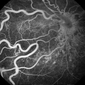
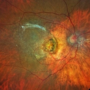 Retinal angiomatous proliferation (RAP)
Jun 15 2022 by Priyanka Raj, MBBS, MS
Retinochoroidal anastomosis seen in stage III Retinal angiomatous proliferation (RAP).
Photographer: Sushil Mishra
Imaging device: Zeiss Clarus 500
Condition/keywords: age-related macular degeneration (AMD), retinal angiomatous proliferation (RAP), retinochoroidal anastomosis
 Loading…