Search results (43 results)
-
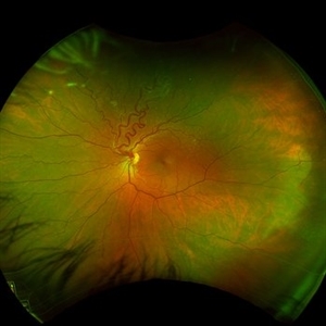
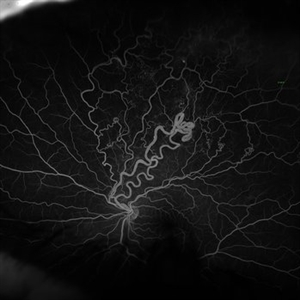
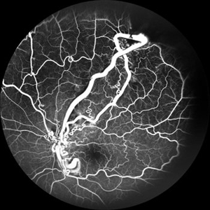 From Artery to Vein, No Detour: Meet the AV Maverick: Racemose Hemangioma
Jul 1 2025 by rohan jain
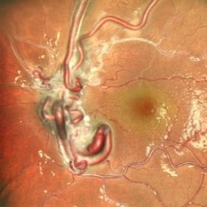
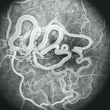
A case of 10 year old girl with defective vision in LE (6/60) who presented us with this condition.
Photographer: Dr. ROHAN JAIN
Imaging device: mirante
Condition/keywords: arteriovenous malformation, FFA in a case of Racemose angioma, racemose hemangioma
-
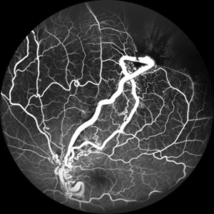 From Artery to Vein, No Detour: Meet the AV Maverick: Racemose Hemangioma
Jul 1 2025 by rohan jain
A case of 10 year old girl with defective vision in LE (6/60) who presented us with this condition.
Photographer: Dr. ROHAN JAIN
Imaging device: mirante
Condition/keywords: arteriovenous malformation, FFA in a case of Racemose angioma, racemose hemangioma
-
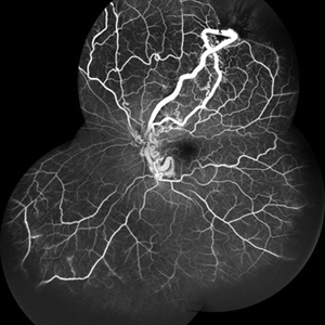 From Artery to Vein, No Detour: Meet the AV Maverick: Racemose Hemangioma
Jul 1 2025 by rohan jain
A case of 10 year old girl with defective vision in LE (6/60) who presented us with this condition.
Photographer: Dr. ROHAN JAIN
Imaging device: mirante
Condition/keywords: arteriovenous malformation, FFA in a case of Racemose angioma, racemose hemangioma
-
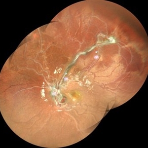 From Artery to Vein, No Detour: Meet the AV Maverick: Racemose Hemangioma
Jul 1 2025 by rohan jain
A case of 10 year old girl with defective vision in LE (6/60) who presented us with this condition.
Photographer: Dr. ROHAN JAIN
Imaging device: mirante
Condition/keywords: arteriovenous malformation, FFA in a case of Racemose angioma, racemose hemangioma
-
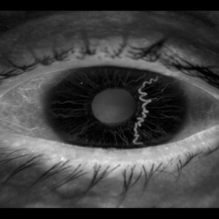 Iris Racemose Hemangioma
Jan 1 2023 by Maxwell J Wingelaar, MD
Fluorescein Angiogram of 66 year old female presented with an iris racemose hemangioma
Photographer: Ken Huff
Condition/keywords: Racemose hemangioma
-
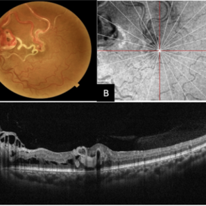 Multimodal Imaging of a Type 3 Retinal Racemose Hemangioma
Sep 8 2024 by Maria Antonia Orrego
We present the case of a 33 year-old woman with visual loss of her left eye since childhood. Fundus examination revealed a retinal arteriovenous malformation with vessels originating from the optic nerve and extending to the fovea and equator, corresponding to a type 3 retinal racemose hemangioma (A). Infrared reflectance imaging confirmed findings described in funduscopy (B). Spectral domain optical coherence tomography shows dilated vessels in the internal and external retinal layers and adjacent intraretinal fluid (C).
Photographer: Dr. Maria Antonia Orrego V, Universidad CES, Clinica Clofán, Medellín, Colombia
Imaging device: Optovue Solix
Condition/keywords: arteriovenous malformation, multimodal imaging, racemose hemangioma, retinal arteriovenous malformations
-
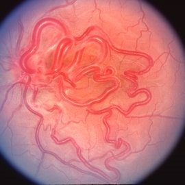
 Racemose Hemangioma
Feb 20 2013 by From the Collections of Thomas M. Aaberg, MD and Thomas M. Aaberg Jr., MD
Dilated tortuous blood vessels.
Condition/keywords: racemose hemangioma
-
 Racemose Hemangioma
Feb 20 2013 by From the Collections of Thomas M. Aaberg, MD and Thomas M. Aaberg Jr., MD
Dilated tortuous blood vessels.
Condition/keywords: racemose hemangioma
-
---thumb.jpg/image-square;max$300,300.ImageHandler) Racemose Hemangioma
Racemose Hemangioma
Feb 20 2013 by From the Collections of Thomas M. Aaberg, MD and Thomas M. Aaberg Jr., MD
dilated vessels
Condition/keywords: racemose hemangioma
-
 Racemose Hemangioma
Dec 16 2025 by Seif Allah Anwar
18 year-old female with dilated, tortuous arteriovenous communication without an intervening capillary bed. Vessels may appear coiled or spaghetti-like extending into the foveal region with no associated retinal hemorrhages, exudates, or edema On OCT : the anomalous vessels appear hyper reflective spanning the whole retinal thickness with ILM draping, No associated subretinal or intraretinal fluid.
Photographer: Seif Anwar , KING SALMAN INTERNATIONAL UNIVERSITY
Imaging device: TOPCON
Condition/keywords: racemose hemangioma
-
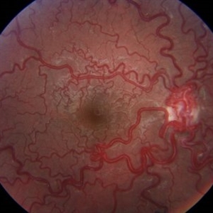
 Racemose Hemangioma
Apr 7 2022 by Sengul Ozdek, MD, FEBO, FASRS
This is a fundus picture of a case with Racemose hemangioma. It is a rare sporadic congenital arteriovenous malformation, with unilateral involvement. It is characterized by a dilated, tortuous, tangled network of retinal vessels, mostly emerging from the optic disc and extending towards periphery, with no distinction between arterioles and venules.
Photographer: Refiye Basdogan
Imaging device: Canon
Condition/keywords: congenital arteriovenous malformation, Racemose hemangioma
-
 Racemose Hemangioma
Mar 14 2021 by Luiz A Zago, PhD
Racemose hemangioma in a 30-year-old woman with ambliopia in this eye.
Photographer: Luiz Alberto Zago Filho
Imaging device: Topcon 50IX
Condition/keywords: racemose hemangioma
-
 Racemose Hemangiomatosis
May 27 2020 by Jamin S. Brown, MD
Fundus photo of 25-year-old female with racemose hemangiomatosis OS.
Photographer: Stefanie Palmer CRA, Retina-Vitreous Surgeons of CNY
Condition/keywords: racemose hemangioma
-
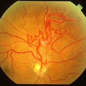
 Retinal Arterio-Venous Malformation / Racemose Angioma
Jan 19 2024 by Brian K Do, MD
Color fundus photo of a 49 year-old woman in whom a vascular abnormality (racemose hemangioma) was noted incidentally during routine ophthalmic examination.
Photographer: Bryan Murphy, The Retina Group of Washington
Condition/keywords: arteriovenous malformation, racemose hemangioma, Wyburn -Mason Syndrome, Wyburn-Mason
-
 Retinal Arterio-Venous Malformation / Racemose Angioma
Jan 19 2024 by Brian K Do, MD
Autofluorescence of the left eye of a 49 year-old woman in whom a vascular abnormality (racemose hemangioma) was noted incidentally during routine ophthalmic examination.
Photographer: Bryan Murphy, The Retina Group of Washington
Condition/keywords: arteriovenous malformation, racemose hemangioma, Wyburn -Mason Syndrome, Wyburn-Mason
-
 Retinal Arterio-Venous Malformation / Racemose Angioma
Jan 19 2024 by Brian K Do, MD
Fluorescein angiogram of a 49 year-old woman in whom a vascular abnormality (racemose hemangioma) was noted incidentally during routine ophthalmic examination.
Photographer: Bryan Murphy, The Retina Group of Washington
Condition/keywords: arteriovenous malformation, racemose hemangioma, Wyburn -Mason Syndrome, Wyburn-Mason
-
 Retinal Arterio-Venous Malformation / Racemose Angioma
Jan 19 2024 by Brian K Do, MD
Fluorescein angiogram of the left eye of a 49 year-old woman in whom a vascular abnormality (racemose hemangioma) was noted incidentally during routine ophthalmic examination.
Photographer: Bryan Murphy, The Retina Group of Washington, Chevy Chase, MD
Condition/keywords: arteriovenous malformation, racemose hemangioma, Wyburn -Mason Syndrome, Wyburn-Mason
-
 Retinal Arteriovenous Malformations (Racemose Hemangiomatosis)
Mar 30 2018 by Rameez N Hussain, MD
A 7-years-old Portuguese girl with unilateral retinal arteriovenous malformations composed of dilated, tortuous vessels with normal vision.
Photographer: Thambi Durai. Consultant Optometrist, Orbit Health Care - Dr Agarwal's Eye Hospital, Maputo, Mozambique
Imaging device: TOPCON
Condition/keywords: racemose hemangioma, retinal arteriovenous malformations, Wyburn-Mason
-
 Retinal Arteriovenous Malformations (Racemose Hemangiomatosis)
Mar 30 2018 by Rameez N Hussain, MD
A 7-year-old Portuguese girl with unilateral retinal arteriovenous malformations composed of dilated, tortuous vessels with normal vision.
Photographer: Thambi Durai, Consultant Optometrist, Orbit Health Care - Dr Agarwal's Eye Hospital, Maputo, Mozambique
Imaging device: TOPCON
Condition/keywords: racemose hemangioma, retinal arteriovenous malformations, Wyburn-Mason
-
---thumb.jpg/image-square;max$300,300.ImageHandler) Retinal AV malformation FA 4
Retinal AV malformation FA 4
Jan 11 2013 by Alex P. Hunyor, MD
Retinal arteriovenous malformation - Archer group 3 - fluorescein angiogram 4 of 4.
Condition/keywords: racemose hemangioma, retinal arteriovenous malformations
-
 Wyburn Mason Syndrome
May 2 2013 by Henry J. Kaplan, MD
Racemose angioma of the retina in Wyburn Mason syndrome.
Condition/keywords: racemose hemangioma
-
 Wyburn-Mason (Raecemose Hemangioma)
Oct 2 2013 by Jerald A. Bovino, MD
The patieint has typical Wyburn Mason syndrome with dilated, extrement tortuous venules.
Condition/keywords: dilated and tortous veins, racemose hemangioma
-
 Wyburn-Mason Syndrome
Apr 8 2019 by Gary R. Cook, MD, FACS
21-year-old white male with congenital arteriovenous communication in his left eye; VA=20/30-2
Condition/keywords: congenital arteriovenous communication of the retina (CAVCR), racemose hemangioma, Wyburn-Mason
-
 Wyburn-Mason Syndrome
Apr 8 2019 by Gary R. Cook, MD, FACS
Fluorescein angiogram image of the 21-year-old white male with congenital arteriovenous communication of the retina (Wyburn-Mason syndrome) OS demonstrating no late leakage of fluorescein dye from the abnormal vessels; V.A. = 20/30-2
Condition/keywords: congenital arteriovenous communication of the retina (CAVCR), fluorescein angiogram (FA), racemose hemangioma, Wyburn-Mason
 Loading…