Search results (41 results)
-
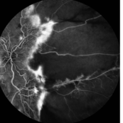
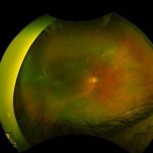
 Active Vasculitis with Proliferative Retinopathy
Jan 30 2021 by Raja Rami P Reddy, MD FRCS FASRS
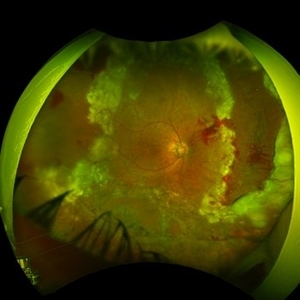
25-year-old boy with unilateral recent onset visual loss. Fundus shows areas of active vasculitis nasally and large neovascular complexes temporally and on the disc and early fibrous membrane formation. Fellow eye fundus is normal. Further investigations suggested tubercular etiology
Photographer: Raja Rami Reddy P
Imaging device: fundus camera
Condition/keywords: proliferative retinopathy, tuberculosis, vasculitis
-
---thumb.jpg/image-square;max$300,300.ImageHandler) Binder3 P12 Slide82
Binder3 P12 Slide82
Feb 15 2013 by From the Collections of Thomas M. Aaberg, MD and Thomas M. Aaberg Jr., MD
Color fundus photograph showing peripheral retinal nonperfusion, retinal neovascularization elsewhere (NVE), venous beading and dilatation, retinal microaneurysms, and intraretinal hemorrhage.
Condition/keywords: peripheral retinal nonperfusion, proliferative retinopathy, retinal neovascularization
-
---thumb.jpg/image-square;max$300,300.ImageHandler) Extensive neovascularization of the disc
Extensive neovascularization of the disc
Feb 15 2013 by From the Collections of Thomas M. Aaberg, MD and Thomas M. Aaberg Jr., MD
Color fundus photograph showing extensive neovascularization of the disc.
Condition/keywords: proliferative retinopathy, retinal neovascularization
-
---thumb.jpg/image-square;max$300,300.ImageHandler) Leakage from peripheral retinal neovascularization and peripheral nonperfusion
Leakage from peripheral retinal neovascularization and peripheral nonperfusion
Feb 15 2013 by From the Collections of Thomas M. Aaberg, MD and Thomas M. Aaberg Jr., MD
Late-phase fluorescein angiograph showing leakage from peripheral retinal neovascularization and peripheral nonperfusion
Condition/keywords: peripheral retinal nonperfusion, proliferative retinopathy, retinal neovascularization
-
---thumb.jpg/image-square;max$300,300.ImageHandler) peripheral retinal nonperfusion, capillary abnormalities, leaking retinal microaneurysms, and blocked fluorescence
peripheral retinal nonperfusion, capillary abnormalities, leaking retinal microaneurysms, and blocked fluorescence
Feb 15 2013 by From the Collections of Thomas M. Aaberg, MD and Thomas M. Aaberg Jr., MD
Mid-phase fluorescein angiograph showing peripheral retinal nonperfusion, capillary abnormalities, leaking retinal microaneurysms, and blocked fluorescence from intraretinal hemorrhage.
Condition/keywords: peripheral retinal nonperfusion, proliferative retinopathy
-
---thumb.jpg/image-square;max$300,300.ImageHandler) Peripheral retinal nonperfusion, capillary abnormalities, retinal microaneurysms, and intraretinal hemorrhage
Peripheral retinal nonperfusion, capillary abnormalities, retinal microaneurysms, and intraretinal hemorrhage
Feb 15 2013 by From the Collections of Thomas M. Aaberg, MD and Thomas M. Aaberg Jr., MD
Color fundus photograph showing peripheral retinal nonperfusion, capillary abnormalities, retinal microaneurysms, and intraretinal hemorrhage.
Condition/keywords: peripheral retinal nonperfusion, proliferative retinopathy
-
---thumb.jpg/image-square;max$300,300.ImageHandler) Peripheral retinal nonperfusion, venous beading and dilatation, retinal microaneurysms, and intraretinal hemorrhage
Peripheral retinal nonperfusion, venous beading and dilatation, retinal microaneurysms, and intraretinal hemorrhage
Feb 15 2013 by From the Collections of Thomas M. Aaberg, MD and Thomas M. Aaberg Jr., MD
Color fundus photograph corresponding to slide titled "staining of retinal vessels, leakage from peripheral retinal neovascularization and peripheral nonperfusion." Shows peripheral retinal nonperfusion, venous beading and dilatation, retinal microaneurysms, and intraretinal hemorrhage.
Condition/keywords: peripheral retinal nonperfusion, proliferative retinopathy, retinal neovascularization
-
 Proliferative Retinopathy
Mar 28 2023 by Harold Rodriguez
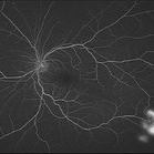
Fluorescein Angiogram on a 43 Year Old female with Proliferative Retinopathy.
Photographer: Harold Rodriguez
Condition/keywords: proliferative retinopathy
-
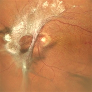 Proliferative Retinopathy
Nov 4 2024 by Tejaswita Verma
Fundus photograph of a middle aged male with diabetes showing large FVP following NVD.
Photographer: DR. TEJASWITA VERMA
Imaging device: MIRANTE
Condition/keywords: FVPs, neovascularization of the disc (NVD), proliferative diabetic retinopathy (PDR)
-
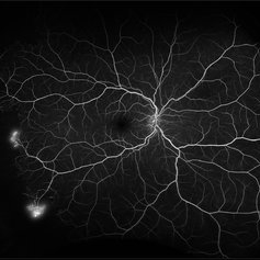
 Proliferative Sickle Cell Retinopathy
Feb 1 2023 by Olivia Rainey
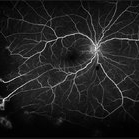
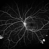
Ultra-widefield fluorescein angiography of a 25-year old male with Proliferative Sickle Cell Retinopathy affecting his left eye. Patient stated that he was born with Sickle disease (SC), and has yearly eye exams. He noted no vision concerns over the last year but has typically experienced sickle attacks about 1-2 per year. The physician noted that the fluorescein obtained showed peripheral nonperfusion affecting the patient's nasal and temporal retina as well as neovascularization affecting his left eye more than his right. He recommended pan retinal photocoagulation in his left eye for his temporal and nasal retina, as as well as his right eye following.
Photographer: Olivia Rainey, OCT-C, COA
Imaging device: Optos California
Condition/keywords: early phase, fluorescein angiogram (FA), fluorescein leakage, left eye, neovascularization (NV), proliferative retinopathy, sickle cell retinopathy, ultra-wide field imaging, ultra-widefield image
-
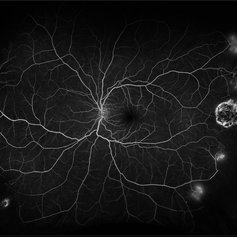
 Proliferative Sickle Cell Retinopathy
Feb 1 2023 by Olivia Rainey
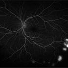
Ultra-widefield fluorescein angiography of a 25-year old male with Proliferative Sickle Cell Retinopathy affecting his right eye. Patient stated that he was born with Sickle disease (SC), and has yearly eye exams. He noted no vision concerns over the last year but has typically experienced sickle attacks about 1-2 per year. The physician noted that the fluorescein obtained showed peripheral nonperfusion affecting the patient's nasal and temporal retina as well as neovascularization affecting his left eye more than his right. He recommended pan retinal photocoagulation in his left eye for his temporal and nasal retina, as as well as his right eye following.
Photographer: Olivia Rainey, OCT-C, COA
Imaging device: Optos California
Condition/keywords: early phase, fluorescein angiogram (FA), fluorescein leakage, neovascularization (NV), non-perfusion, proliferative retinopathy, right eye, sickle cell retinopathy, ultra-wide field imaging, ultra-widefield image
-
 Proliferative Sickle Cell Retinopathy, Color OD
May 23 2018 by Hosam Attia, MD
45-year-old African American, male with sickle cell anemia (SC disease) with arteriolar attenuation, mild venous tortuosity, Sunburst (S) and large, partially auto-infarcted sea fan with fresh heme, OD.
Imaging device: Optos California Ultra-Wide Field Fundus Camera
Condition/keywords: neovascularization elsewhere (NVE), proliferative retinopathy, sea fan, sickle cell, sickle cell retinopathy
-
 Proliferative Sickle Cell Retinopathy, Color OD
May 23 2018 by Hosam Attia, MD
45-year-old African American, male with sickle cell anemia (SC disease) with arteriolar attenuation, mild venous tortuosity, Sunburst (S) and large, partially auto-infarcted Seafan with fresh heme, OD.
Imaging device: Optos California Ultra-Wide Field Fundus Camera
Condition/keywords: neovascularization elsewhere (NVE), proliferative retinopathy, sea fan, sickle cell, sickle cell retinopathy
-
 Proliferative Sickle Cell Retinopathy, Color OS
May 23 2018 by Hosam Attia, MD
45-year-old African American, male with sickle cell anemia (SC disease ) with arteriolar attenuation, mild venous tortuosity, peripheral arterio-venous anastomoses (shown better on red free), multiple small NVEs/ early sea fans (one w/ early auto-infarction) and sunburst (S) - (Not showing very well in photos) OS.
Imaging device: Optos California Ultra-Wide Field Fundus Camera
Condition/keywords: neovascularization elsewhere (NVE), proliferative retinopathy, sea fan, sickle cell, sickle cell retinopathy
-
 Proliferative Sickle Cell Retinopathy, Early phase FA OD
May 23 2018 by Hosam Attia, MD
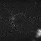
Fluorescein angiogram photograph of a 45-year-old African American, male with sickle cell anemia (SC disease), depicting extensive peripheral capillary non-perfusion, with early hyperfluorescence over the ischemic retina temporally, with late staining and diffuse leakage consistent with partially auto-infarcted, but active NVE/sea fan OD.
Imaging device: Optos California Ultra-Wide Field Fundus Camera
Condition/keywords: neovascularization elsewhere (NVE), proliferative retinopathy, sea fan, sickle cell, sickle cell retinopathy
-
 Proliferative Sickle Cell Retinopathy, Early phase FA OS
May 23 2018 by Hosam Attia, MD
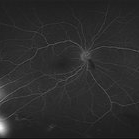
Fluorescein angiogram photograph of a 45-year-old African American, male with cell anemia (SC disease ), depicting peripheral capillary non-perfusion, with multiple, small area of early to mid phase hyperfluorescence over the ischemic retina temporally, with mild late leakage consistent with active NVEs/ early sea fans OS.
Imaging device: Optos California Ultra-Wide Field Fundus Camera
Condition/keywords: neovascularization elsewhere (NVE), proliferative retinopathy, sea fan, sickle cell, sickle cell retinopathy
-
 Proliferative Sickle Cell Retinopathy, Late FA OS
May 23 2018 by Hosam Attia, MD
Fluorescein angiogram photograph of a 45-year-old African American, male with sickle cell anemia (SC disease), depicting peripheral capillary non-perfusion, with multiple, small area of mild late leakage consistent with active NVEs/ early Seafans OS.
Imaging device: Optos California Ultra-Wide Field Fundus Camera
Condition/keywords: neovascularization elsewhere (NVE), proliferative retinopathy, sea fan, sickle cell, sickle cell retinopathy
-
 Proliferative Sickle Cell Retinopathy, Late phase FA OD
May 23 2018 by Hosam Attia, MD
Fluorescein angiogram photograph of a 45-year-old African American, male with sickle cell anemia (SC disease), depicting extensive peripheral capillary non-perfusion, with late staining and diffuse leakage consistent with partially auto-infarcted, but active NVE/sea fan OD.
Imaging device: Optos California Ultra-Wide Field Fundus Camera
Condition/keywords: neovascularization elsewhere (NVE), proliferative retinopathy, sea fan, sickle cell, sickle cell retinopathy
-
 Proliferative Sickle Cell Retinopathy, Mid phase FA OS
May 23 2018 by Hosam Attia, MD
Fluorescein angiogram photograph of a 45-year-old African American, male with sickle cell anemia (SC disease), depicting peripheral capillary non-perfusion, with multiple, small area of early to mid phase hyperfluorescence over the ischemic retina temporally, with mild late leakage consistent with active NVEs/ early sea fans OS.
Imaging device: Optos California Ultra-Wide Field Fundus Camera
Condition/keywords: neovascularization elsewhere (NVE), proliferative retinopathy, sea fan, sickle cell, sickle cell retinopathy
-
 Proliferative Sickle Cell Retinopathy, Red Free OD
May 23 2018 by Hosam Attia, MD
Red free fundus photo of a 45-year-old African American, male with sickle cell anemia (SC Disease ) with arteriolar attenuation, mild venous tortuosity, Sunburst (S) and large, partially auto-infarcted sea fan, OD.
Imaging device: Optos California Ultra-Wide Field Fundus Camera
Condition/keywords: neovascularization elsewhere (NVE), proliferative retinopathy, sea fan, sickle cell, sickle cell retinopathy
-
 Proliferative Sickle Cell Retinopathy, Red Free OS
May 23 2018 by Hosam Attia, MD
Red free fundus photograph of a 45-year-old African American, male with sickle cell anemia (SC disease) with arteriolar attenuation, mild venous tortuosity, peripheral arterio-venous anastomoses (Inferotemporally), multiple small NVEs/ early sea fans OS.
Photographer: Aaron Appiah, M.D.
Imaging device: Optos California Ultra-Wide Field Fundus Camera
Condition/keywords: neovascularization elsewhere (NVE), proliferative retinopathy, sea fan, sickle cell, sickle cell retinopathy
-
 Proliferative Sickle Retinopathy
Sep 4 2018 by John S. King, MD
Initial picture before Dr. Hruby performed laser. Peripheral ischemia and some "sea fan" retinal neovascularization present.
Photographer: Pam Hall
Imaging device: Optos CA
Condition/keywords: proliferative retinopathy, sickle cell retinopathy
-
 Proliferative Sickle Retinopathy
Sep 4 2018 by John S. King, MD
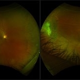
Initial photo (left) and 4 months after Dr. Hruby performed peripheral circumferential retinal scatter photocoagulation to zone of peripheral ischemia (right). Regression of neovascularization.
Photographer: Stacey
Imaging device: Optos CA
Condition/keywords: laser photocoagulation, proliferative retinopathy, sickle cell retinopathy
-
 Retinal Detachment Following Scleral Buckling, Retinectomy, Laser, and Oil
Jan 31 2022 by Ahmad B. Tarabishy, MD
Ultra wide-field fundus photograph of a 55-year-old gentleman who is 4 days after surgery with scleral buckling, pars plana vitrectomy, perfluoron tamponade, membrane peeling, direct fluid-PFO-oil exchange, nasal and temporal retinectomies, and endolaser photocoagulation. Visual acuity was 20/150 under oil.
Photographer: Megan McLandsborough, Lakeland Eye Clinic
Imaging device: Optos California UWF Camera
Condition/keywords: endolaser, Membrane Peel, PPV, proliferative retinopathy, proliferative vitreoretinopathy (PVR), Retinal Detachment, retinal detachment with retinal defect, scleral buckle, submacular perfluorocarbon liquid (PFO)
-
 Retinal Detachment with Proliferative Vitreoretinopathy
Jan 31 2022 by Ahmad B. Tarabishy, MD
Ultra wide-field fundus photograph of a 55-year-old gentleman who had previously underwent laser retinopexy for multiple inferior retinal breaks. He presented with a macula-off retinal detachment from a new temporal break with proliferative vitreoretinopathy with fixed folds noted temporally and superonasally.
Photographer: Megan McLandsborough, Lakeland Eye Clinic
Imaging device: Optos California UWF Camera
Condition/keywords: laser retinopexy, macula off Retinal Detachment, proliferative retinopathy, proliferative vitreoretinopathy (PVR), Retinal Detachment, retinal detachment with retinal defect
 Loading…