Search results (493 results)
-
 "Boat-Shaped" Preretinal Hemorrhage
Feb 21 2019 by Mitzy E Torres Soriano, MD
Color fundus photograph showing preretinal (subhyaloid) hemorrhage in a diabetic patient with proliferative diabetic retinopathy.
Photographer: Andrea Vitale, MD
Condition/keywords: preretinal hemorrhage, proliferative diabetic retinopathy (PDR), subhyaloid hemorrhage
-
 A Fleet of Boat-Shaped Hemorrhages
Aug 1 2024 by James P Dossett, MD
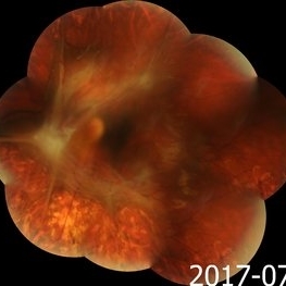
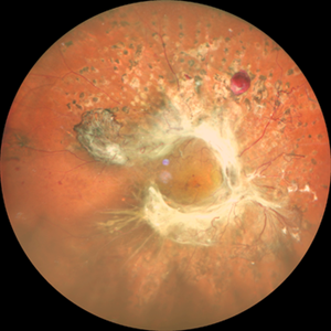
Pseudocolor fundus photograph of the left eye of a 54-year-old diabetic man presenting with bilateral vision loss. Examination revealed 20/200 vision OS with extensive preretinal and vitreous hemorrhage, marked diffuse neovascularization, macular edema and hard exudates.
Photographer: Beth Smith, West Virginia University Eye Institute
Condition/keywords: proliferative diabetic retinopathy (PDR)
-
 Active diabetic retinopathy despite PRP
Oct 30 2022 by Diego Andrés Rodriguez, MD
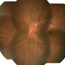
A 52-year-old patient with active proliferative diabetic retinopathy despite good glycemic control and PRP performed 1 year ago in the right eye
Photographer: Sociedad de Cirugía Ocular
Imaging device: Clarus 700
Condition/keywords: diabetic retinopathy, pan-retinal photocoagulation (PRP), proliferative diabetic retinopathy (PDR), wide angle imaging
-
 Active Laser Modified Proliferative Diabetic Retinopathy
Mar 17 2024 by Hector Gabriel Moreno Solano, MD, MHA
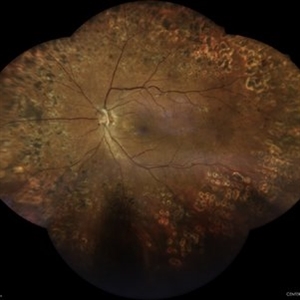
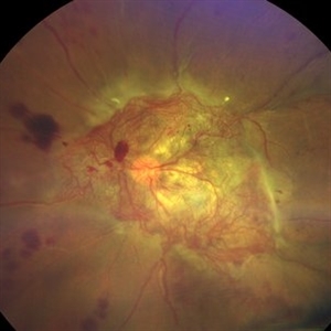
Wide-field composite fundus images of a 78-year-old male patient with a single functional eye due to poorly controlled diabetic retinopathy with activity data and multiple photocoagulation traces.
Photographer: Héctor Gabriel Moreno-Solano, MD, MHA
Imaging device: Centervue Eidon
Condition/keywords: Diabetes, proliferative diabetic retinopathy (PDR), retinopathy
-
 Active neovascularization in Proliferative Diabetic Retinopathy
Jan 10 2018 by Peter H. Tang, MD, PhD
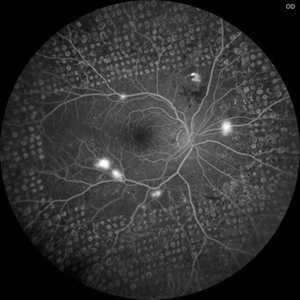
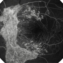
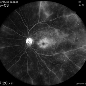
Fluorescein angiography image from a 46-year-old woman with uncontrolled proliferative diabetic retinopathy shows extensive dye leakage from active neovascularization.
Imaging device: Optos California
Condition/keywords: diabetes, diabetic retinopathy, fluorescein leakage, neovascularization elsewhere (NVE), neovascularization of the disc (NVD), pan-retinal photocoagulation (PRP), proliferative diabetic retinopathy (PDR)
-
 Active Proliferative Diabetic Retinopathy
Jul 12 2024 by Korey Starkey
Fluorescein angiogram performed on 35 year old female with active proliferative diabetic retinopathy. Patient has peripapillary vascular loop and history of PRP treatment in both eyes. Patients left eye vision measured at Dcc20/200-1 at this visit.
Photographer: Korey Starkey
Imaging device: Optos
Condition/keywords: FLUORESCEIN ANGIOGRAPHY, hyperfluorescence, laser scarring, Optos, proliferative diabetic retinopathy (PDR), sea fan, ultra-wide field imaging, vascular loop
-
 Active Proliferative Diabetic Retinopathy
Aug 16 2022 by Donnie Willis
51 yo female. Uncontrolled Diabetes. Active Proliferative Diabetic Retinopathy.
Photographer: Donnie Willis, Tennessee Retina
Imaging device: Optos
Condition/keywords: capillary dropouts, Diabetes, fluorescein angiogram (FA), OPTOS, proliferative diabetic retinopathy (PDR), tractional retinal detachment
-
 Advanced PDR
Sep 1 2014 by Hamid Ahmadieh, MD
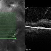
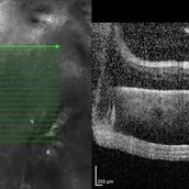
OCT image of the right eye of a 50-year-old woman with advanced PDR.
Photographer: Soodabeh Fooladian, Negah Eye Center, Tehran, Iran
Condition/keywords: optical coherence tomography (OCT), proliferative diabetic retinopathy (PDR)
-
 Advanced PDR
Sep 1 2014 by Hamid Ahmadieh, MD
OCT image of the left eye of a 50-year-old woman with advanced PDR.
Photographer: Soodabeh Fooladian, Negah Eye Center, Tehran, Iran
Condition/keywords: optical coherence tomography (OCT), proliferative diabetic retinopathy (PDR)
-
 Advanced PDR
Sep 1 2014 by Hamid Ahmadieh, MD
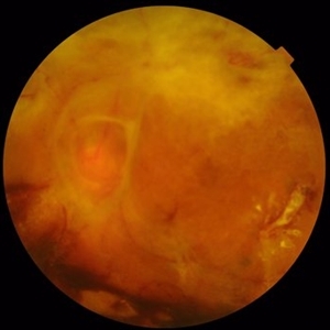
Color fundus photograph of the left eye of a 50-year-old woman with advanced PDR.
Photographer: Soodabeh Fooladian, Negah Eye Center, Tehran, Iran
Condition/keywords: color fundus photograph, proliferative diabetic retinopathy (PDR)
-
 Advanced PDR
Sep 1 2014 by Hamid Ahmadieh, MD
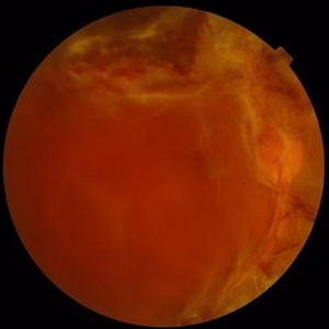
Color fundus photograph of the right eye of a 50-year-old woman with advanced PDR.
Photographer: Soodabeh Fooladian, Negah Eye Center, Tehran, Iran
Condition/keywords: color fundus photograph, proliferative diabetic retinopathy (PDR), subhyaloid hemorrhage
-
 Advanced PDR Left Eye
Aug 31 2014 by Neha Goel, MS DNB FRCS (Glasg)
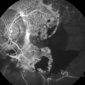
Fundus photograph of the left eye.
Photographer: Neha Goel
Imaging device: Zeiss Visucam
Condition/keywords: fibrovascular proliferation, ischaemic diabetic maculopathy, proliferative diabetic retinopathy (PDR)
-
 Advanced PDR Left Eye FFA
Aug 31 2014 by Neha Goel, MS DNB FRCS (Glasg)
Fluorescein angiogram of the left eye.
Photographer: Neha Goel
Imaging device: Zeiss Visucam
Condition/keywords: fibrovascular proliferation, ischaemic diabetic maculopathy, proliferative diabetic retinopathy (PDR)
-
 Advanced PDR RE FFA
Aug 31 2014 by Neha Goel, MS DNB FRCS (Glasg)
Fluorescein angiogram of the right eye.
Photographer: Neha Goel
Imaging device: Zeiss Visucam
Condition/keywords: fibrovascular proliferation, ischaemic diabetic maculopathy, proliferative diabetic retinopathy (PDR)
-
 Advanced PDR-RE
Aug 31 2014 by Neha Goel, MS DNB FRCS (Glasg)
Fundus photograph of the right eye of a 50-year-old diabetic male.
Photographer: Neha Goel
Imaging device: Zeiss Visucam
Condition/keywords: fibrovascular proliferation, ischaemic diabetic maculopathy, proliferative diabetic retinopathy (PDR)
-
 Advanced Proliferative Diabetic Retinopathy
Nov 4 2017 by Hamid Ahmadieh, MD
Merged color fundus photograph of the left eye of a 30-year-old woman with type1 diabetes since childhood. Note laser scars, severe fibrous proliferation, traction RD and macular dragging.
Photographer: Shabnam Poureh, Negah Eye Center, Tehran, Iran
Condition/keywords: color fundus photograph, diabetes, fibrous proliferation, proliferative diabetic retinopathy (PDR), severe traction
-
 Advanced Proliferative Diabetic Retinopathy With Fibrovascular Proliferation
Jan 4 2019 by Isha Agarwalla
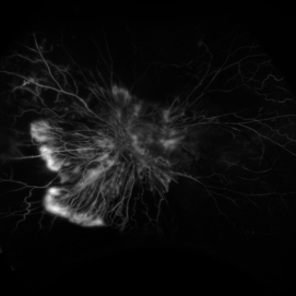
A 29-year-old female with a long-standing history of diabetes mellitus presented with a fibrovascular membrane (FVM) at the viteroretinal interface due to underlying inflammation and angiogenesis induced by ischemia. FVM involved the disc and extended towards the superior and inferior arcades along with extensive capillary drop out areas due to micro aneurysms.
Condition/keywords: fibrovascular proliferation, proliferative diabetic retinopathy (PDR)
-
 Advanced Proliferative Diabetic Retinopathy With Fibrovascular Proliferation
Jan 4 2019 by Isha Agarwalla
A 29-year-old female with a long-standing history of diabetes mellitus presented with a fibrovascular membrane(FVM) at the viteroretinal interface due to underlying inflammation and angiogenesis induced by ischemia. FVM involved the disc and extended towards the superior and inferior arcades along with extensive capillary drop out areas due to micro aneurysms.
Condition/keywords: fibrovascular proliferation, fluorescein angiogram (FA), proliferative diabetic retinopathy (PDR)
-
 An Intricate Web of Vasculature
Jan 5 2022 by SHISHIR VERGHESE, MS, FVRS, FAICO (Retina)
Fundus photograph of a 55-year-old gentleman with decreased vision in the left eye for 6 months. History of uncontrolled diabetes and hypertension for 15 years. Best corrected visual acuity in the left eye was 5/60.
Photographer: SHISHIR VERGHESE
Imaging device: ZEISS CLARUS
Condition/keywords: diabetes, proliferative diabetic retinopathy (PDR)
-
 Angiographic Diabetic Macular Edema in a Case of Proliferative Diabetic Retinopathy
Apr 9 2024 by Akansha Sharma
Fundus fluorescein angiographic image of 62 year old male demonstrating angiographic diabetic macular edema in a case of proliferative diabetic retinopathy.
Photographer: Dr. Akansha Sharma, Bharati Eye Hospital
Condition/keywords: clinically significant macular edema (CSME), diabetic blindness, diabetic macular edema, proliferative diabetic retinopathy (PDR)
-
 Annular Tractional Retinal Detachment
Jul 4 2024 by Hector Gabriel Moreno Solano, MD, MHA
52-year-old Hispanic female patient with a diagnosis of type II diabetes mellitus of 15 years of evolution, comes to the retina service for progressive visual loss in the right eye (single functional eye) with visual acuity of 20/100, Fundus examination reveals laser-modified proliferative diabetic retinopathy with activity + annular tractional retinal detachment with macular involvement.
Photographer: Hector Gabriel Moreno Solano, MD, MHA, HGZ #20 IMSS Puebla.
Imaging device: Mirante
Condition/keywords: macular detachment, proliferative diabetic retinopathy (PDR), tractional retinal detachment
-
 Aurora Borealis in Retina
Apr 25 2025 by Poornachandra B, MS, FVRS
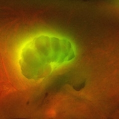
Fundus picture of 54 year old male with proliferative diabetic retinopathy with fluorescent blood clot in vitreous cavity.
Photographer: Mr Dhikshith
Imaging device: Optos daytona
Condition/keywords: blood, proliferative diabetic retinopathy (PDR)
-
 Before and After Vitrectomy
Nov 17 2023 by Bradley T. Smith, MD, FASRS
Middle age male diabetic retinopathy and resolving exudate following repair of tractional detachment with membrane peeling.
Condition/keywords: coats-like response, Diabetes, fibrotic neovascularization, fibrovascular proliferation, pars plana vitrectomy (PPV), proliferative diabetic retinopathy (PDR), tractional retinal detachment
-
 Bilateral CRVO and PDR
Nov 4 2021 by Stefanie Palmer
Patient with both PDR and CRVO, 34 year old female, post-COVID.
Photographer: Stefanie Palmer, CRA
Imaging device: Topcon
Condition/keywords: central retinal vein occlusion (CRVO), COVID-19, diabetic retinopathy, proliferative diabetic retinopathy (PDR), venous beading
 Loading…