Search results (66 results)
-
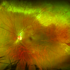
 "Boat-Shaped" Preretinal Hemorrhage
Feb 21 2019 by Mitzy E Torres Soriano, MD
Color fundus photograph showing preretinal (subhyaloid) hemorrhage in a diabetic patient with proliferative diabetic retinopathy.
Photographer: Andrea Vitale, MD
Condition/keywords: preretinal hemorrhage, proliferative diabetic retinopathy (PDR), subhyaloid hemorrhage
-
---thumb.JPG/image-square;max$300,300.ImageHandler) Acute myeloid leukemia
Acute myeloid leukemia
Dec 9 2012 by Mallika Goyal, MD
Right eye of a 21-year-old gentleman with acute myeloid leukemia who is undergoing chemotherapy and has low platelet counts (17,000) shows multiple pre-retinal haemorrhages. Other eye has similar picture. There is no vascular occlusion or inflammation. Visual prognosis remains good with spontaneous resolution expected over few weeks.
Photographer: Mallika Goyal, MD, Apollo Health City, Hyderabad, India
Condition/keywords: preretinal hemorrhage
-
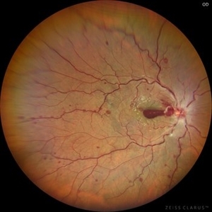 Central Retinal Vein Occlusion with Preretinal Hemorrhage
Mar 16 2021 by MOHIT GUPTA
Fundus photograph of right eye of a young male after 2nd dose of Covishield vaccine presented to us with central retinal vein occlusion and preretinal hemorrhage at macula in right eye.
Photographer: Dr Mohit Gupta , Prakash Netra Kendr, Lucknow, India
Imaging device: zeiss clarus
Condition/keywords: central retinal vein occlusion (CRVO), preretinal hemorrhage
-
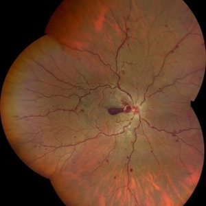 CRVO With Preretinal Hemorrhage
Mar 16 2021 by MOHIT GUPTA
Fundus photograph of right eye of a young male after 2nd dose of Covishield vaccine presented to us with central retinal vein occlusion and preretinal hemorrhage at macula in right eye.
Photographer: Dr Mohit Gupta
Imaging device: Zeiss Clarius
Condition/keywords: central retinal vein occlusion (CRVO), preretinal hemorrhage
-
 Dengue Fever
Oct 25 2012 by Mallika Goyal, MD
Fundus photograph of the right eye of a 32-year-old gentleman with dengue fever and thrombocytopenia. Photograph shows extensive retinal and pre-retinal haemorrhages, roth spots but no dengue retinitis. Same patient as in images 1-5.
Condition/keywords: Dengue Fever, preretinal hemorrhage, rosacea conjunctivitis
-
 Dengue Fever
Oct 25 2012 by Mallika Goyal, MD
Fundus photograph of the left eye of a 32-year-old gentleman with dengue fever and thrombocytopenia. Photograph shows extensive retinal and pre-retinal haemorrhages, roth spots but no dengue retinitis. Same patient as in images 1-5.
Condition/keywords: Dengue Fever, preretinal hemorrhage, rosacea conjunctivitis
-
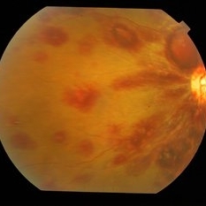 Dengue Fever
Oct 25 2012 by Mallika Goyal, MD
Fundus photograph of the left eye of a 32-year-old gentleman with dengue fever and thrombocytopenia. Photograph shows extensive retinal and pre-retinal haemorrhages, roth spots but no dengue retinitis. Same patient as in images 1-5
Condition/keywords: Dengue Fever, preretinal hemorrhage, rosacea conjunctivitis
-
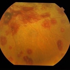 Dengue Fever
Oct 25 2012 by Mallika Goyal, MD
Fundus photograph of the left eye of a 32-year-old gentleman with dengue fever and thrombocytopenia. Photograph shows extensive retinal and pre-retinal haemorrhages, roth spots but no dengue retinitis.
Condition/keywords: Dengue Fever, preretinal hemorrhage, rosacea conjunctivitis
-
 Dense Preretinal Hemorrhage
May 15 2020 by Iuri Golubev, MD
34-year-old male w/h/o DM type 1 and PDR.
Condition/keywords: preretinal hemorrhage, proliferative diabetic retinopathy (PDR)
-
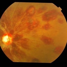
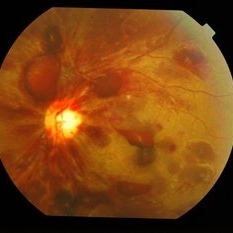
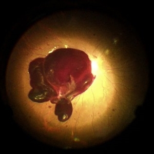
 High risk Proliferative Diabetic Retinopathy treated with Pan Retinal Photocoagulation
Nov 5 2022 by Somnath Chakraborty, MD
A Fundus Photo Montage of 43 year old Asian Male with Type 2 Diabetes Mellitus since 7 years who presented with sudden onset diminition of vision in his Left eye. BCVA OS was 20/200. He was diagnosed to have Pre retinal bleed due to Proliferative Diabetic Retinopathy and was treated with Pan Retinal Photocoagulation. This image shows a large neo-cascular frond at the disc and superior to it with Pre-retinal bleed and Fresh laser marks along
Photographer: Pulak Roy
Condition/keywords: diabetic blindness, diabetic retinopathy vitrectomy study (DRVS), fresh laser burns, laser photocoagulation, preretinal hemorrhage, proliferative diabetic retinopathy (PDR)
-
 Laser Photocoagulation
Nov 9 2012 by Norman Byer
This shows the same lesion four days after laser photo coagulation. The new hemorrhages seen in this photograph did not occur during the photocoagulation but developed within the next four days.
Condition/keywords: argon photocoagulation, laser photocoagulation, preretinal hemorrhage
-
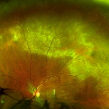
 Macroaneurysm
Jun 23 2021 by Cláudia Farinha
Color Optomap from a middle-aged man with preretinal hemorrhage due to a macroaneurysm.
Photographer: Claudia Farinha, MD
Imaging device: Optomap, Optos
Condition/keywords: macroaneurysm, preretinal hemorrhage
-
 Massive Commotio Retinae
Oct 20 2020 by Veronika Yehezkeli
24-year-old man was injured from an explosion of a plastic bottle towards the nasal conjunctiva of his left eye. A massive commotio retinae was diagnosed superotemporally.
Photographer: Veronika Yehezkeli, Meir medical center, Israel
Condition/keywords: blunt trauma, commotio retinae, preretinal hemorrhage
-
 Massive Commotio Retinae
Oct 20 2020 by Veronika Yehezkeli
Fundus photograph of a 24-year-old male, made after blunt trauma with a plastic bottle. Note massive commotio retinae and preretinal hemorrhages in the contralateral to trauma area.
Photographer: Veronika Yehezkeli, Meir medical center, Israel
Condition/keywords: blunt trauma, commotio retinae, preretinal hemorrhage, trauma
-
 PDR with Active NVD
Oct 8 2012 by Jeffrey G. Gross, MD, FASRS
PDR with active NVD and preretinal hemorrhage, mild VH and partial PRP.
Condition/keywords: neovascularization of the disc (NVD), preretinal hemorrhage, scatter laser photocoagulation, vitreous hemorrhage
-
 PDR with Active NVD
Oct 8 2012 by Jeffrey G. Gross, MD, FASRS
PDR with active NVD and preretinal hemorrhage.
Condition/keywords: neovascularization of the disc (NVD), preretinal hemorrhage
-
 Post-Op Day 10 Barrier Laser
Feb 13 2013 by From the Collections of Thomas M. Aaberg, MD and Thomas M. Aaberg Jr., MD
Barrier laser, preretinal hemorrhage.
Condition/keywords: barrier laser, post-op, preretinal hemorrhage
-
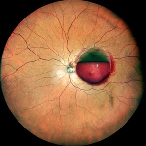
 Pre Macular Subhyaloid Hemorrhage
Jan 20 2021 by Nivesh Gupta
A 41 year old male patient complaining of diminution of vision in left eye since 6 days. His best corrected visual acuity finger counting at 2 meters.
Photographer: Nivesh Gupta, Retina Fellow, Retina Foundation, Ahmedabad, India
Condition/keywords: hypertensive retinopathy, preretinal hemorrhage, subhyaloid hemorrhage
-
---thumb.jpg/image-square;max$300,300.ImageHandler) Pre-Retinal Fibrous Proliferative Membrane
Pre-Retinal Fibrous Proliferative Membrane
Feb 20 2013 by From the Collections of Thomas M. Aaberg, MD and Thomas M. Aaberg Jr., MD
Color photo of various views on the fundus and focused on the midvitreous level.
Condition/keywords: color photo, preretinal hemorrhage
-
 Pre-Retinal Hemorrhage With Disc Edema
Apr 19 2024 by Akansha Sharma
Color fundus photograph of a 39 year old female with disc edema along with a pre-retinal hemorrhage.
Photographer: Dr. Akansha Sharma, Bharati Eye Hospital
Condition/keywords: disc edema, preretinal hemorrhage
-
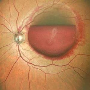
 Premacular Subhyaloid Hemorrhage
Jan 20 2021 by Nivesh Gupta
A 41-year-old male patient complaining of diminution of vision in left eye since 6 days. His best corrected visual acuity finger counting at 2 meters.
Photographer: Nivesh Gupta, Retina Fellow, Retina Foundation, Ahmedabad, India
Condition/keywords: hypertensive retinopathy, preretinal hemorrhage, subhyaloid hemorrhage
-
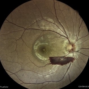
 Preretinal Bleed in Lasered PDR
Jun 4 2014 by Neha Goel, MS DNB FRCS (Glasg)
Fundus photograph of the right eye of a 40-year-old diabetic male. Panretinal photocoagulation had been performed. There is a dense preretinal bleed overlying the macula and nasal to the disc.
Photographer: Neha Goel
Imaging device: Zeiss Visucam
Condition/keywords: laser photocoagulation, neovascularization elsewhere (NVE), preretinal hemorrhage, proliferative diabetic retinopathy (PDR)
-
 Preretinal Hemorrhage
May 6 2017 by Mitzy E Torres Soriano, MD
Fundus photograph of a 36-year-old-woman with a preretinal subhyaloid hemorrhage (valsalva retinopathy).
Photographer: Mitzy Torres Soriano
Condition/keywords: macular hemorrhage, premacular hemorrhage, preretinal hemorrhage, subhyaloid hemorrhage, valsalva retinopathy
-
 Preretinal Hemorrhage
Sep 20 2012 by Allen Chiang, MD, FASRS
34-year old woman with preretinal hemorrhage in the macula, with dehemoglobinization occuring within the central portion of the hemorrhage while undergoing observation.
Imaging device: Zeiss Cirrus
Condition/keywords: preretinal hemorrhage
 Loading…