Search results (30 results)
-
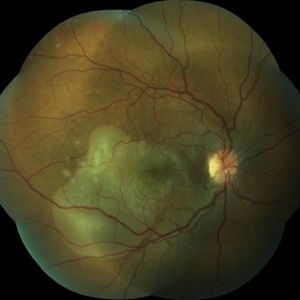
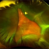 Choroidal Detachment
Oct 4 2018 by Emily Cooper
Optos photograph of an 80-year-old man presenting with red, painful eye after heart surgery.
Photographer: Emily Cooper, Retina Specialists of Michigan, Grand Rapids MI
Imaging device: Optos
Condition/keywords: choroidal detachment, posterior scleritis
-
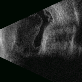 Posterior Nodular Scleritis
Apr 23 2025 by Gustavo Uriel Fonseca Aguirre
This B-mode ultrasound scan demonstrates a posterior scleral nodule accompanied by vitritis, serous retinal detachment, and annular choroidal detachment. The nodule appears as a localized hypoechoic scleral thickening, while the serous retinal detachment shows a smooth convex configuration. The choroidal detachment presents with the characteristic ring-shaped elevation, suggesting significant intraocular inflammation or hypotony.
Photographer: Gustavo U. Fonseca Aguirre, Hospital Conde de Valenciana, Ciudad de México
Condition/keywords: posterior nodular scleritis, posterior scleritis
-
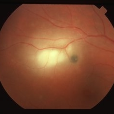
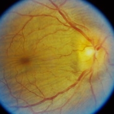 Posterior Scleritis
Sep 12 2023 by Ben Serar
Fundus photograph of RE showing Disc edema with Choroidal folds in a case of Posterior Scleritis.
Condition/keywords: chorioretinal folds, disc edema, Posterior scleritis
-
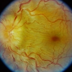 Posterior Scleritis
Sep 12 2023 by Ben Serar
Fundus photograph of LE showing Disc edema with Choroidal folds in a case of Posterior Scleritis
Condition/keywords: chorioretinal folds, disc edema, posterior scleritis
-
 POSTERIOR SCLERITIS
Nov 1 2023 by ANKIT JAIN
USG B SACN image showing typical T-sign in axial horizontal view with increased thickening of the sclero-choroidal complex suggestive of posterior scleritis
Photographer: DR ANKIT JAIN
Condition/keywords: B scan ultrasound, posterior scleritis, ULTRASOUND
-
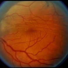
 Posterior Scleritis
Jan 4 2025 by Tejaswita Verma
Left eye fundus photo of a 49 year old female presenting with 5 days history of blurred vision, Vision was finger counting 2 mts. and nodular posterior scleritis with T sign on USG was present. OCT revealed altered foveal contour with septations and SRF pockets and bacillary layer detachment.
Photographer: DR. TEJASWITA VERMA
Imaging device: MIRANTE
Condition/keywords: posterior scleritis
-
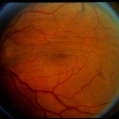
 Posterior Scleritis
Aug 5 2024 by Aniruddh Soni, DO DNB FLVPEI
Fundus photo of a 35 year old female presenting with pain and defective vision.
Photographer: Dr Aniruddh Soni
Imaging device: Zeiss Visucam 500
Condition/keywords: posterior scleritis
-
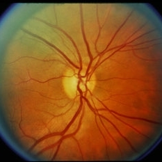
 Posterior Scleritis
Apr 2 2019 by Gary R. Cook, MD, FACS
White female with a focus of acute posterior scleritis beneath the superotemporal arcade OD; VA = 20/40-2
Imaging device: Topcon VT-50
Condition/keywords: posterior scleritis
-
 Posterior Scleritis
Jun 6 2019 by Gary R. Cook, MD, FACS
Mid-phase fluorescein angiogram image of acute posterior scleritis lesion beneath the superotemporal arcade OD; V.A. = 20/40-2
Imaging device: Topcon VT-50
Condition/keywords: FA mid phase, fluorescein angiogram (FA), posterior scleritis
-
 Posterior Scleritis
Nov 18 2013 by Mallika Goyal, MD
Subretinal fluid in a 30-year-old lady with posterior scleritis. This resolved with intravenous pulsed steroids for 3 days.
Photographer: Mallika Goyal, MD, Apollo Health City, Hyderabad
Condition/keywords: posterior scleritis
-
 Posterior Scleritis
Dec 8 2013 by Mallika Goyal, MD
Rapid onset serous macular detachment in a young lady with scleritis. Treated with pulsed intravenous steroids with resolution.
Photographer: Mallika Goyal, MD, Apollo Health City, Hyderabad, India
Condition/keywords: posterior scleritis
-
 Posterior Scleritis
Dec 8 2013 by Mallika Goyal, MD
Resolving fluid at macula 1 week after pulsed intravenous steroids for posterior scleritis with serous macular detachment in a young lady.
Photographer: Mallika Goyal, MD, Apollo Health City, Hyderabad, India
Condition/keywords: posterior scleritis
-
 Posterior Scleritis
Dec 8 2013 by Mallika Goyal, MD
Flat macula 3 weeks after pulsed intravenous steroids for posterior scleritis with serous macular detachment in a young lady.
Photographer: Mallika Goyal, MD, Apollo Health City, Hyderabad, India
Condition/keywords: posterior scleritis
-
---thumb.JPG/image-square;max$300,300.ImageHandler) Posterior Scleritis Atypical
Posterior Scleritis Atypical
Dec 13 2013 by Mallika Goyal, MD
Right eye fundus of a 32-year-old male presenting with unilateral reduced quality of vision, pain and headache for 5 days; visual acuity was 20/25. There was trace RAPD, white conjunctiva, no intraocular inflammation, mild disc edema and congestion, normal retina and macula. OCT was normal. A diagnosis of optic neuritis was considered, later revised to posterior scleritis with contiguous papillitis.
Photographer: Mallika Goyal, MD, Apollo Health City, Hyderabad, India
Condition/keywords: posterior scleritis
-
---thumb.JPG/image-square;max$300,300.ImageHandler) Posterior Scleritis Atypical
Posterior Scleritis Atypical
Dec 13 2013 by Mallika Goyal, MD
Normal left eye fundus of a 32-year-old male presenting with right eye posterior scleritis with contiguous papillitis
Photographer: Mallika Goyal, MD, Apollo Health City, Hyderabad, India
Condition/keywords: posterior scleritis
-
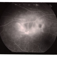
---thumb.JPG/image-square;max$300,300.ImageHandler) Posterior Scleritis Atypical
Posterior Scleritis Atypical
Dec 13 2013 by Mallika Goyal, MD
Right eye early phase fluorescein angiogram of a 32-year-old male with posterior scleritis with contiguous papillitis shows early peripapillary hyperfluorescence suggestive of generalised inflammation in posterior pole.
Photographer: Mallika Goyal, MD, Apollo Health City, Hyderabad, India
Condition/keywords: posterior scleritis
-
---thumb.JPG/image-square;max$300,300.ImageHandler) Posterior Scleritis Atypical
Posterior Scleritis Atypical
Dec 13 2013 by Mallika Goyal, MD
Right eye late phase fluorescein angiogram of a 32-year-old male with posterior scleritis with contiguous papillitis shows staining of disc with peripapillary hyperfluorescence suggestive of generalised inflammation in posterior pole.
Photographer: Mallika Goyal, MD, Apollo Health City, Hyderabad, India
Condition/keywords: posterior scleritis
-
---thumb.JPG/image-square;max$300,300.ImageHandler) Posterior Scleritis Atypical
Posterior Scleritis Atypical
Dec 13 2013 by Mallika Goyal, MD
Left eye fluorescein angiogram of a 32-year-old male with fellow eye posterior scleritis shows normal angiogram in contrast to the disc staining and peripapillary hyperfluorescence seen in the affected right eye.
Photographer: Mallika Goyal, MD, Apollo Health City, Hyderabad, India
Condition/keywords: posterior scleritis
-
---thumb.JPG/image-square;max$300,300.ImageHandler) Posterior Scleritis Atypical
Posterior Scleritis Atypical
Dec 13 2013 by Mallika Goyal, MD
Right eye fluorescein angiogram of a 32-year-old male with posterior scleritis with contiguous papillitis shows staining of disc with peripapillary hyperfluorescence suggestive of generalized inflammation in posterior pole.
Photographer: Mallika Goyal, MD, Apollo Health City, Hyderabad, India
Condition/keywords: posterior scleritis
-
---thumb.JPG/image-square;max$300,300.ImageHandler) Posterior Scleritis Atypical
Posterior Scleritis Atypical
Dec 13 2013 by Mallika Goyal, MD
Right eye fluorescein angiogram of a 32-year-old male with posterior scleritis with contiguous papillitis shows staining of disc with peripapillary hyperfluorescence suggestive of generalised inflammation in posterior pole.
Photographer: Mallika Goyal, MD, Apollo Health City, Hyderabad, India
Condition/keywords: posterior scleritis
-
---thumb.JPG/image-square;max$300,300.ImageHandler) Posterior Scleritis Atypical
Posterior Scleritis Atypical
Dec 13 2013 by Mallika Goyal, MD
Right eye fundus of a 32-year-old male presenting with unilateral reduced quality of vision, pain and headache for 5 days; visual acuity was 20/25. There was trace RAPD, white conjunctiva, no intraocular inflammation, mild disc edema and congestion, normal retina and macula. OCT was normal. A diagnosis of optic neuritis was considered, later revised to posterior scleritis with contiguous papillitis.
Photographer: Mallika Goyal, MD, Apollo Health City, Hyderabad, India
Condition/keywords: posterior scleritis
-
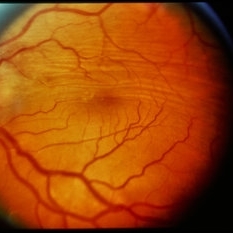
 Posterior Scleritis (Focal)
Sep 10 2014 by David Callanan, MD
47-white female, posterior scleritis (focal).
Condition/keywords: scleritis
-
 Posterior Scleritis (Focal)
Sep 10 2014 by David Callanan, MD
47-white female, posterior scleritis (focal).
Condition/keywords: scleritis
-
 Posterior Scleritis (Focal)
Sep 10 2014 by David Callanan, MD
47-white female, posterior scleritis (focal).
Condition/keywords: scleritis
-
 Posterior Scleritis (Focal)
Sep 10 2014 by David Callanan, MD
47-white female, posterior scleritis (focal).
Condition/keywords: scleritis
 Loading…