Search results (120 results)
-
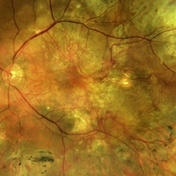
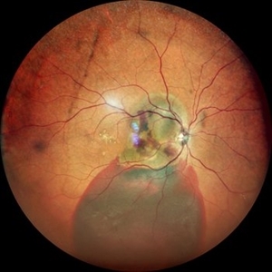
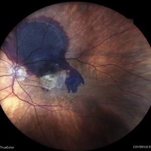
 Branching vascular network..Polypoidal choroidal vasculopathy
Feb 21 2022 by Shobhit Chawla, M.S.
A 56 year old patient on treatment with ANTIVGEF therapy for six years
Photographer: Shobhit Chawla
Imaging device: Zeiss Clarus 500
Condition/keywords: branching vascular network (BVN), polypoidal choroidal vasculopathy (PCV)
-
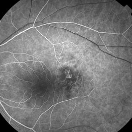
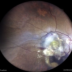
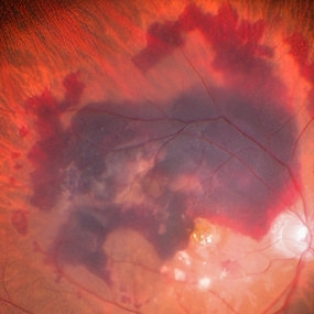
 BVN Branching Vascular Network in PCV
Feb 19 2022 by Vishal Gupta, MBBS, MS
Very evident branching vascular network in the fellow eye of one eyed patient who lost the other eye to massive hemorrhagic PCV in a 47 year old male patient.
Photographer: Dr Shobhit Chawla, Prakash Netra Kendr, Lucknow, UP, INDIA
Imaging device: Zeiss Clarus 500
Condition/keywords: branching vascular network (BVN), polypoidal choroidal vasculopathy (PCV)
-
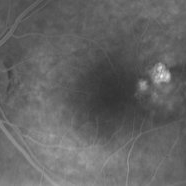
 Choroidal Nevus with PCV
Jan 31 2018 by John S. King, MD
16 sec
Imaging device: topcon
Condition/keywords: choroidal neovascular membrane (CNVM), choroidal nevus, polypoidal choroidal vasculopathy (PCV)
-
 Choroidal Nevus with PCV
Jan 31 2018 by John S. King, MD
28 sec
Imaging device: topcon
Condition/keywords: choroidal neovascular membrane (CNVM), polypoidal choroidal vasculopathy (PCV)
-
 Choroidal Nevus with PCV
Jan 31 2018 by John S. King, MD
2:12
Imaging device: topcon
Condition/keywords: choroidal neovascular membrane (CNVM), choroidal nevus, polypoidal choroidal vasculopathy (PCV)
-
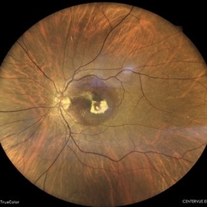
 Choroidal Nevus with PCV
Jan 31 2018 by John S. King, MD
44-year-old AAF without syptoms; nevus, rpe alterations, few exudates at base of polyp.
Imaging device: Topcon
Condition/keywords: choroidal neovascular membrane (CNVM), choroidal nevus, polypoidal choroidal vasculopathy (PCV)
-
 Choroidal nevus with polypoidal choroidal vasculopathy
Nov 20 2012 by Roy Schwartz, MD
Rare combination of a choroidal nevus complicated by polypoidal choroidal vasculopathy. The lesion is temporal to the fovea.
Photographer: Galit Yair-Pur
Condition/keywords: choroidal nevus, polypoidal choroidal vasculopathy (PCV)
-
 choroidal nevus with polypoidal choroidal vasculopathy
Nov 20 2012 by Roy Schwartz, MD
Rare combination of a choroidal nevus complicated by polypoidal choroidal vasculopathy. The lesion is temporal to the fovea, and leakage of subretinal fluid almost reaching the fovea is demonstrated.
Photographer: Galit Yair-Pur
Condition/keywords: choroidal nevus, polypoidal choroidal vasculopathy (PCV)
-
 choroidal nevus with polypoidal choroidal vasculopathy
Nov 20 2012 by Roy Schwartz, MD
Rare combination of a choroidal nevus complicated by polypoidal choroidal vasculopathy. The lesion is temporal to the fovea, and subretinal fluid almost reaching the fovea is demonstrated.
Photographer: Galit Yair-Pur
Condition/keywords: choroidal nevus, optical coherence tomography (OCT), polypoidal choroidal vasculopathy (PCV)
-
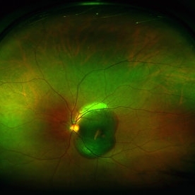
 Fundus Photo of IPCV
Aug 10 2019 by Manish Nagpal, MD, FRCS (UK), FASRS
Fundus showing serosanguinous collection under macular suggestive of active IPCV lesion.
Photographer: Gayathri Mohan, Retina Foundation
Imaging device: Nidek Mirante SLO
Condition/keywords: polypoidal choroidal vasculopathy (PCV)
-
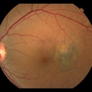
 Idiopathic Polypoidal Choroidal Vasculopathy
May 11 2022 by Pramod Kumar Suman, MBBS, MD
Fundus photograph of an 59-year-old male with clinically visible orange-red sub-retinal nodules with sub-retinal hemorrhage.
Photographer: Pramod Kumar Suman, Retina Foundation, Ahmedabad
Condition/keywords: polypoidal choroidal vasculopathy (PCV)
-
 Idiopathic Polypoidal Choroidal Vasculopathy
Mar 26 2024 by Akansha Sharma
Color fundus photograph of a 61 year old treatment naive female patient with scarring at fovea with surrounding subretinal bleed suggestive of idiopathic polypoidal choroidal vasculopathy.
Photographer: Dr. Akansha Sharma, Bharati Eye Hospital
Condition/keywords: polypoidal choroidal vasculopathy (PCV)
-
 Idiopathic Polypoidal Choroidal Vasculopathy
Apr 9 2024 by Akansha Sharma
Color fundus photograph of a 74 year old female with subretinal bleed in a case of polypoidal choroidal vasculopathy.
Photographer: Dr. Akansha Sharma, Bharati Eye Hospital
Condition/keywords: PCV, polypoidal choroidal vasculopathy (PCV), wet age-related macular degeneration (wet AMD)
-
 IDIOPATHIC POLYPOIDAL CHOROIDAL VASCULOPATHY
Jun 6 2023 by Akansha Sharma
COLOUR FUNDUS PHOTOGRAPH OF AN 80 YEAR OLD MALE PATIENT WITH IDIOPATHIC POLYPOIDAL CHOROIDAL VASCULOPATHY
Photographer: Dr. Urmil Shah, Dr. Denish Patel, Dr. Akansha Sharma
Condition/keywords: idiopathic polypoidal choroidal vasculopathy, polypoidal choroidal vasculopathy (PCV)
-
 Large Subretinal Bleed in Case of Wet ARMD
Sep 28 2024 by Anjana Mirajkar, MS Ophthalmology
An intra operative image showing large sub retinal hemorrhage involving the macular area and along the superior arcade with exudation at the macular area in case of wet ARMD.
Photographer: Dr. Anjana Mirajkar -Retina Foundation, Ahmedabad
Condition/keywords: polypoidal choroidal vasculopathy (PCV), subretinal hemorrhage, wet age-related macular degeneration (wet AMD)
-
 Massive Submacular Hemorrhage
Aug 3 2013 by Yusuke Oshima, MD, PhD
Wide-angle viewing photography of a 67-year-old man with massive submacular hemorrhage.
Condition/keywords: polypoidal choroidal vasculopathy (PCV), submacular hemorrhage, subretinal hemorrhage
-
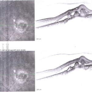
 Neovascular AMD with Polypoidal Choroidal Vasculopathy
Jun 11 2024 by Gregg T. Kokame, MD, MMM, FASRS
Structure OCT from the OCT angiography shows definition of the PCV complex.
Photographer: Jaclyn Pisano
Condition/keywords: branching vascular network (BVN), OCTA, polypoidal choroidal vasculopathy (PCV), wet age-related macular degeneration (wet AMD)
-
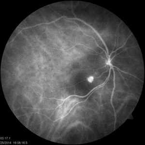
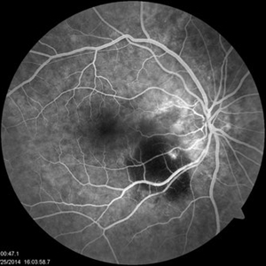
 PCV
PCV
Jul 1 2014 by John S. King, MD
OCT post-PDT. Middle age Mediterranean female with acute decrease in vision mainly characterized as paracentral scotoma. Suboptimal response to short course of anti-VEGF; responded well to PDT.
Photographer: Wayne A Ladlee Jr
Imaging device: Cirrus
Condition/keywords: polypoidal choroidal vasculopathy (PCV)
-
 PCV
PCV
Jul 1 2014 by John S. King, MD
This ICG taken at same time as FA. Middle age Mediterranean female with acute decrease in vision mainly characterized as paracentral scotoma. Suboptimal response to short course of anti-VEGF; responded well to PDT.
Photographer: Wayne A Ladlee Jr
Imaging device: ICG
Condition/keywords: polypoidal choroidal vasculopathy (PCV)
-
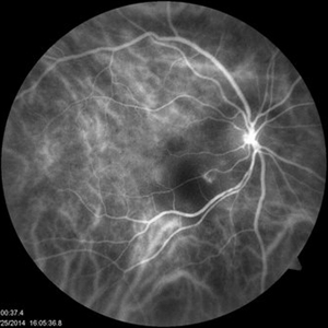
 PCV
PCV
Jul 1 2014 by John S. King, MD
This FA taken after at least one Avastin. Middle age Mediterranean female with acute decrease in vision mainly characterized as paracentral scotoma. Suboptimal response to short course of anti-VEGF; responded well to PDT.
Photographer: Wayne A Ladlee Jr
Imaging device: FA
Condition/keywords: polypoidal choroidal vasculopathy (PCV)
-
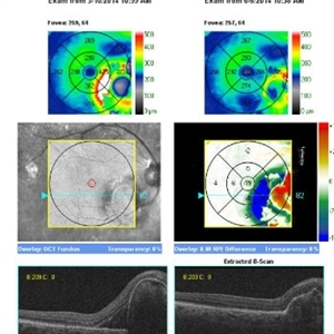
 PCV
PCV
Jul 1 2014 by John S. King, MD
OCT post anti-VEGF. Middle age Mediterranean female with acute decrease in vision mainly characterized as paracentral scotoma. Suboptimal response to short course of anti-VEGF; responded well to PDT.
Photographer: Wayne A Ladlee Jr
Imaging device: Cirrus
Condition/keywords: polypoidal choroidal vasculopathy (PCV)
-
 PCV
PCV
Jul 1 2014 by John S. King, MD
OCT compares presentation to post-PDT. Middle age Mediterranean female with acute decrease in vision mainly characterized as paracentral scotoma. Suboptimal response to short course of anti-VEGF; responded well to PDT.
Photographer: Wayne A Ladlee Jr
Imaging device: Cirrus
Condition/keywords: polypoidal choroidal vasculopathy (PCV)
-
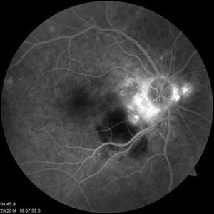
 PCV
PCV
Jul 1 2014 by John S. King, MD
This ICG taken at same time as FA. Middle age Mediterranean female with acute decrease in vision mainly characterized as paracentral scotoma. Suboptimal response to short course of anti-VEGF; responded well to PDT.
Photographer: Wayne A Ladlee Jr
Imaging device: ICG
Condition/keywords: polypoidal choroidal vasculopathy (PCV)
-
 PCV
PCV
Jul 1 2014 by John S. King, MD
This FA taken after at least one Avastin. Middle age Mediterranean female with acute decrease in vision mainly characterized as paracentral scotoma. Suboptimal response to short course of anti-VEGF; responded well to PDT.
Photographer: Wayne A Ladlee Jr
Imaging device: FA
Condition/keywords: polypoidal choroidal vasculopathy (PCV)
-
 PCV
PCV
Jul 1 2014 by John S. King, MD
Photo taken after at least one Avastin. Middle age Mediterranean female with acute decrease in vision mainly characterized as paracentral scotoma. Suboptimal response to short course of anti-VEGF; responded well to PDT.
Photographer: Wayne A Ladlee Jr
Condition/keywords: polypoidal choroidal vasculopathy (PCV)
 Loading…