Search results (21 results)
-
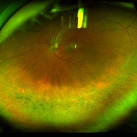
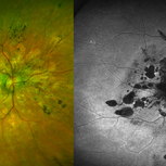
 Branch Retinal Vein Occlusion With Peripheral Pigmentary Change
Jan 15 2019 by Olivia Rainey
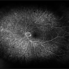
Ultra-wide field fluorescein angiogram of an 85-year-old female with a branch retinal vein occlusion with peripheral pigmentary changes. Patient developed a BRVO after a PPV for an epiretinal membrane.
Photographer: Olivia Rainey
Imaging device: Optos
Condition/keywords: branch retinal vein occlusion (BRVO), epiretinal membrane (ERM), fluorescein angiogram (FA), left eye, Optos, pigmentary retinal dystrophy
-
 Cone-Rod Dystrophy
Jul 20 2023 by Harsh Vardhan Singh, MS
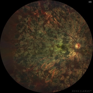
52-year-old male with a advanced stage of cone-rod dystrophy
Photographer: Harsh Vardhan Singh, AIIMS, Guwahati
Imaging device: Zeiss Clarus 700
Condition/keywords: cone dystrophy, Cone-Rod Dystrophy, pigmentary retinal dystrophy, retinal dystrophy
-
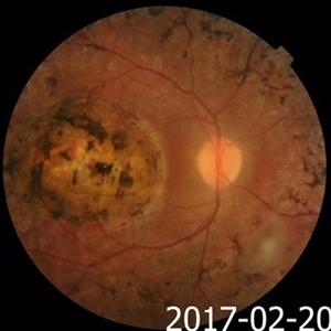
 Macular Coloboma and Pigmentary Retinopathy
Feb 25 2017 by Hamid Ahmadieh, MD
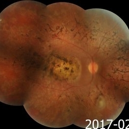
Merged color fundus photograph of the right eye of a 25-year-old woman with the history of low vision since childhood. Bilateral macular colobomata and pigmentary retinopathy similar to Leber's congenital amaurosis are present.
Photographer: Shabnam Poureh, Negah Eye Center, Tehran, Iran
Condition/keywords: bilateral pigmentary retinopathy, color fundus photograph, macular coloboma, pigmentary retinal dystrophy
-
 Macular Coloboma and Pigmentary Retinopathy
Feb 25 2017 by Hamid Ahmadieh, MD
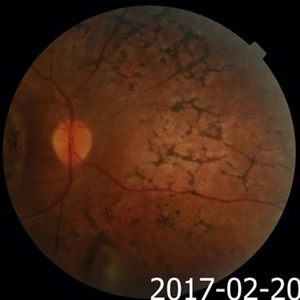
Color fundus photograph of the right eye of a 25-year-old woman with the history of low vision since childhood. Bilateral macular colobomata and pigmentary retinopathy similar to Leber's congenital amaurosis are present.
Photographer: Shabnam Poureh, Negah Eye Center, Tehran, Iran
Condition/keywords: color fundus photograph, pigmentary retinal dystrophy
-
 Macular Coloboma and Pigmentary Retinopathy
Feb 25 2017 by Hamid Ahmadieh, MD
Color fundus photograph of the right eye of a 25-year-old woman with the history of low vision since childhood. Bilateral macular colobomata and pigmentary retinopathy similar to Leber's congenital amaurosis are present.
Photographer: Shabnam Poureh, Negah Eye Center, Tehran, Iran
Condition/keywords: bilateral pigmentary retinopathy, color fundus photograph, macular coloboma, pigmentary retinal dystrophy
-
 Pigmentary Retinal Dystrophy
May 5 2020 by Olivia Rainey
Ultra-widefield fundus autofluorescence image of an 44-year-old male with pigmentary retinal dystrophy affecting both eyes. He presented with decreased night vision for 6 months prior to his appointment. He stated that his recovery time from transitioning from dark to light areas is reduced. He stated that his peripheral vision has never been very good for most of his life. He admits to environmental hearing loss. Patient denies family history of blin. His vision was 20/20 in both eyes. His full field ERG, visual fields were not consistent with RP. Genetic testing with ID Your IRD and annual follow up has been recommended.
Photographer: Olivia Rainey, OCT-C, COA
Imaging device: Optos California
Condition/keywords: fundus autofluorescence (FAF), hyperautofluorescence, hypoautofluorescence, inferior retina, left eye, Optos, ultra-wide field imaging
-
 Pigmentary Retinal Dystrophy
May 5 2020 by Olivia Rainey
Ultra-widefield pseudocolor image of an 44-year-old male with pigmentary retinal dystrophy affecting both eyes. He presented with decreased night vision for 6 months prior to his appointment. He stated that his recovery time from transitioning from dark to light areas is reduced. He stated that his peripheral vision has never been very good for most of his life. He admits to environmental hearing loss. Patient denies family history of blin. His vision was 20/20 in both eyes. His full field ERG, visual fields were not consistent with RP. Genetic testing with ID Your IRD and annual follow up has been recommended.
Photographer: Olivia Rainey, OCT-C, COA
Imaging device: Optos California
Condition/keywords: inferior retina, left eye, Optos, pigment, pseudocolor, ultra-wide field imaging
-
 Pigmentary Retinal Dystrophy
May 5 2020 by Olivia Rainey
Ultra-widefield fundus autofluorescence image of an 44-year-old male with pigmentary retinal dystrophy affecting both eyes. He presented with decreased night vision for 6 months prior to his appointment. He stated that his recovery time from transitioning from dark to light areas is reduced. He stated that his peripheral vision has never been very good for most of his life. He admits to environmental hearing loss. Patient denies family history of blin. His vision was 20/20 in both eyes. His full field ERG, visual fields were not consistent with RP. Genetic testing with ID Your IRD and annual follow up has been recommended.
Photographer: Olivia Rainey, OCT-C, COA
Imaging device: Optos California
Condition/keywords: fundus autofluorescence (FAF), hyperautofluorescence, hypoautofluorescence, inferior retina, Optos, pigment, ultra-wide field imaging
-
 Pigmentary Retinal Dystrophy
May 5 2020 by Olivia Rainey
Ultra-widefield pseudocolor image of an 44-year-old male with pigmentary retinal dystrophy affecting both eyes. He presented with decreased night vision for 6 months prior to his appointment. He stated that his recovery time from transitioning from dark to light areas is reduced. He stated that his peripheral vision has never been very good for most of his life. He admits to environmental hearing loss. Patient denies family history of blin. His vision was 20/20 in both eyes. His full field ERG, visual fields were not consistent with RP. Genetic testing with ID Your IRD and annual follow up has been recommended.
Photographer: Olivia Rainey, OCT-C, COA
Imaging device: Optos California
Condition/keywords: inferior retina, Optos, pigmentary retinal dystrophy, pseudocolor, ultra-wide field imaging
-
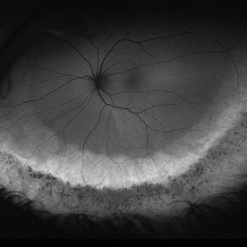
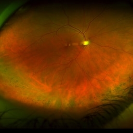
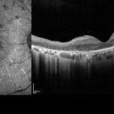
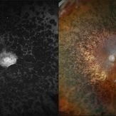
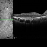 Pigmentary Retinal Dystrophy
Mar 29 2019 by Jessica Norkus
Heidelberg Spectralis image of 41-year-old male patient with pigmentary retinal dystrophy. Atypical findings due to unilateral presentation. Patient has been experiencing symptoms for 15 years, notes significant nyctalopia.
Photographer: Jessica Norkus
Imaging device: Heidelberg Spectralis
Condition/keywords: bone spicule, Heidelburg Spectralis, optical coherence tomography (OCT), pigment changes, unilateral blindness
-
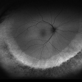
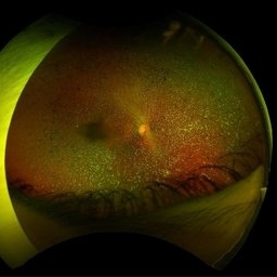
 Pigmentary Retinal Dystrophy
Mar 29 2019 by Jessica Norkus
Optos ultra wide field image of 41-year-old male patient with pigmentary retinal dystrophy. Atypical findings due to unilateral presentation. Patient has been experiencing symptoms for 15 years, notes significant nyctalopia.
Photographer: Jessica Norkus
Imaging device: Optos Ultra Wide Field Camera
Condition/keywords: abnormal fundus, bone spicule, color fundus photograph, color photo, fundus autofluorescence (FAF), fundus photograph, Optos, peripheral bone spicules, pigment changes, ultra-wide field imaging, unilateral blindness
-
 Pigmentary Retinal Dystrophy
Mar 29 2019 by Jessica Norkus
Optos ultra wide field image of 41-year-old male patient with pigmentary retinal dystrophy. Atypical findings due to unilateral presentation. Patient has been experiencing symptoms for 15 years, notes significant nyctalopia.
Photographer: Jessica Norkus
Imaging device: Optos Ultra Wide Field Camera
Condition/keywords: abnormal fundus, bone spicule, color fundus photograph, color photo, fundus photograph, Optos, peripheral bone spicules, pigment changes, ultra-wide field imaging, unilateral blindness
-
 Pigmentary Retinal Dystrophy
Jul 18 2025 by Kimberly Wakester
Optomap RGB and AF of the left eye of an 76-year-old woman with pigmentary retinal dystrophy. No progression of the bone spicules noted on exam and optos imaging. Will continue yearly follow care with dilated exam and optos imaging.
Photographer: Kimberly Wakester, COA, OCT-C
Imaging device: Optos California
Condition/keywords: pigmentary retinal dystrophy
-
 Pigmentary Retinal Dystrophy
Oct 30 2025 by Kimberly Wakester
Optomap RGB of an 77-year-old-woman with Pigmentary Retinal Dystrophy in the left eye. Patient is to continue follow up care yearly with dilated exam and diagnostic testing.
Photographer: Kimberly Wakester, COA, OCT-C
Imaging device: Optos California
Condition/keywords: bone spicules, Pigmentary Retinal Dystrophy
-
 Pigmentary Retinal Dystrophy
Feb 9 2015 by Matt Poe, COA
This was a lady that presented with bilateral pigmentary retinal dystrophy.
Photographer: Matt Poe, COA. Northwest Arkansas Retina Associates, Springdale, AR.
Condition/keywords: hereditary retinal dystrophy, pigmentary retinal dystrophy
-
---thumb.jpg/image-square;max$300,300.ImageHandler) Retinal Dystrophy
Retinal Dystrophy
Aug 9 2013 by From the Collections of Thomas M. Aaberg, MD and Thomas M. Aaberg Jr., MD
Pigmented dystrophy.
Condition/keywords: pigmentary retinal dystrophy, retinal dystrophy
-
---thumb.jpg/image-square;max$300,300.ImageHandler) Retinal Dystrophy
Retinal Dystrophy
Aug 9 2013 by From the Collections of Thomas M. Aaberg, MD and Thomas M. Aaberg Jr., MD
Pigmented dystrophy.
Condition/keywords: pigmentary retinal dystrophy, retinal dystrophy
-
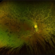
 Retinitis Pigmentosa
Apr 30 2015 by Mitzy E Torres Soriano, MD
Fundus of patient with retinitis pigments, bone spicule-shaped pigment deposits are present with retinal atrophy, while the macula is preserved . Retinal vessels are attenuated.
Photographer: Mitzy E. Torres Soriano, MD; Centro medico Cagua-Estado Aragua. Venezuela
Imaging device: TRC-NW8
Condition/keywords: pigmentary retinal dystrophy, retinal dystrophy, retinitis pigmentosa, retinitis pigmentosa (RP) dystrophy
-
 Retinitis Pigmentosa Associated with Asteroid Hyalosis
Jul 21 2023 by Mohammadkarim Johari
Fundus photograph of an 43-year-old lady with pigmentary retinal dystrophy and asteroid hyalosis, also shadow of posterior subcapsular cataract is obvious.
Photographer: Mohammadkarim Johari, Shiraz university of medical science
Condition/keywords: asteroid hyalosis, pigmentary retinal dystrophy, retinitis pigmentosa (RP) dystrophy
-
 Severe macular atrophy secondary to Pseudoxanthoma Elasticum and CNV
Jul 26 2019 by Olivia Rainey
Ultra-wide field color/autofluorescence comparison of a 54-year-old female with severe macular atrophy secondary to pseudoxanthoma elasticum. Patient developed choroidal neovascularization that does not warrant treatment due to the patient's poor visual acuity. Patient has significantly attenuated ERG. Genetic testing is recommended to rule out retinitis pigmentosa. The mechanism is yet to be determined, but there is a thought that ABCC6 mutation causes alteration of plasma lipid composition may be implicated in the systemic changes observed in PXE.
Photographer: Olivia Rainey
Imaging device: Optos
Condition/keywords: angioid streaks, atrophic central lesion, autofluorescence imaging, choroidal neovascularization (CNV), hypofluorescent lesions, macular atrophy, Optos, pigmentary retinal dystrophy, pseudoxanthoma elasticum (PXE), ultra-wide field imaging
 Loading…