Search results (3 results)
-
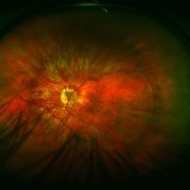 Color Fundus Photograph of Myope With PVD and Staphyloma
Jun 11 2016 by Philip J. Polkinghorne, MD
Color photograph of patient with PVD and staphyloma.
Imaging device: Optos
Condition/keywords: degenerative myopia, peripheral vascular disease (PVD), staphyloma
-
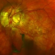 Morning-Glory-Syndrome
Dec 22 2017 by James B. Soque, CRA, OCT-C, COA, FOPS
68-year-old WM with Morning Glory Syndrome with PVD OS with Staphyloma surrounding optic nerve and extending into the macula. Also, esotropia OS from V1 nerve paresis from birth, with amblyopia.
Photographer: James B Soque, CRA OCT-C COA FOPS
Imaging device: Optos Daytona
Condition/keywords: color photo, esotropia, fundus photograph, Optomap, Optos, peripheral vascular disease (PVD), posterior vitreous detachment, staphyloma, ultra-wide field imaging, wide angle imaging
-
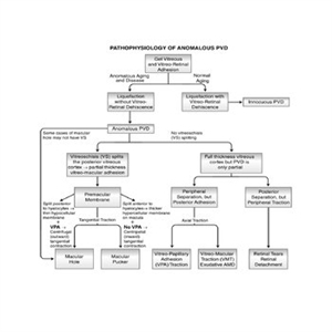 Pathophysiology of Anomalous PVD
Sep 1 2020 by J. Sebag, MD, FACS, FRCOphth, FARVO
This unifying concept of vitreo-retinopathies hypothesizes that the pathogenesis of several vitreoretinal diseases that were previously considered very disparate, are actually all manifestations of the same underlying pathophysiology – anomalous PVD. Note that vitreo-papillary adhesion (VPA) and traction can cause primary optic neuropathy, but might also play a role in facilitating/promoting cell migration and proliferation during pathologic neovascularization of the optic disc. Further, VPA seems to alter the vector of tangential forces exerted by a membrane, in some cases full-thickness posterior vitreous cortex and in some cases the outer layer of the posterior vitreous cortex left attached to the macula after vitreoschisis. While not all cases of macular holes have vitreoschisis, they feature vitreomacular adhesion and traction almost always with VPA. [From Sebag J: Anomalous PVD – a unifying concept in vitreo-retinal diseases. Graefe’s Arch Clin Exp Ophthalmol 2004;242:690-8 and Sebag J, Niemeyer M, Koss M: Anomalous PVD and vitreoschisis. In: Vitreous – in Health & Disease (J. Sebag, ed.) Springer, New York, 2014, pg. 252; image © Springer Nature, reprinted with permission]
Condition/keywords: pathology, peripheral vascular disease (PVD)
 Loading…