Search results (6 results)
-
 Peripheral Drusen
-
 Peripheral Drusen
-
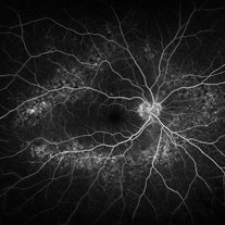 Peripheral Drusen
Jan 19 2022 by Olivia Rainey
Ultra-widefield fluorescein angiogram of an 83-year-old female with Peripheral Drusen affecting both eyes. The patient presented on 1/19/2022 with slightly decreased vision since her last appointment. Her vision was sc20/25-2 in the right eye. The physician did not believe that the peripheral drusen represents AMD and recommended monitoring. The patient also had mild diabetic retinopathy at the time of her visit.
Photographer: Olivia Rainey, OCT-C, COA
Imaging device: Optos California
Condition/keywords: background diabetic retinopathy (BDR), fluorescein angiogram (FA), Optos, Peripheral drusen, staining, ultra-wide field imaging
-
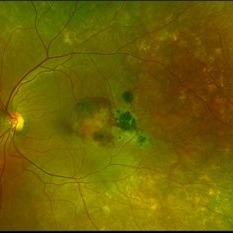 Subretinal Hemorrhage with Chorioretinal Macular Scars
Sep 28 2022 by Denica Rodriguez
Ultra-widefield pseudocolor fundus photograph of a 59 year old female with Subretinal Hemorrhage with Chorioretinal Macular Scars affecting her left eye. The physician presumes the etiology is CNV from adjacent scarring/choroidal rupture. Patient has history of ocular trauma with cricket ball at age 10-12 years old. She suspects that she might have suffered choroidal rupture, which has resulted in secondary CNV and hemorrhage that we are seeing today. She recommends treatment with Eylea sample injection in a series of 4 at a 4-5 week interval. The patient's vision at the time of her appointment was Dcc20/40-2 PHNI.
Photographer: Denica Rodriguez, COA
Imaging device: Optos California
Condition/keywords: antiVEGF therapy, chorioretinal scar, choroidal neovascular membrane (CNVM), fundus photography, left eye, macular scar, Optos, peripheral drusen, pseudocolor, secondary CNV, subretinal hemorrhage, ULTRA WIDE FIELD, ultra-wide field imaging
-
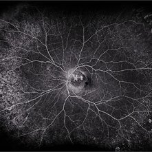
 Midperipheral Drusen
Dec 18 2014 by H. Michael Lambert, MD
Midperipheral drusen OS in a 15-year-old white female with peau d'orange fundus changes but biopsy negative for PXE.
Condition/keywords: drusen, peau d'orange fundus
-
 PPCNVM and Peripheral Drusen Seen on Optos FA
Apr 22 2020 by John S. King, MD
72-year-old white male c/o of distortion OS for about 2 months. 20/100 OS, normotensive, small grey-green subretinal area just temporal to the optic disc. FA shows leakage c/w a ppcnvm; there is some SR and IR leakage as well as staining of peripheral drusen and some window defects from cobblestone. Avastin was adminstered.
Photographer: Asli Ahmed
Imaging device: CA
Condition/keywords: drusen, peripapillary choroidal neovascularization (PPCNVM)
 Loading…