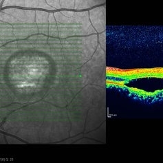
Search results (308 results)
-
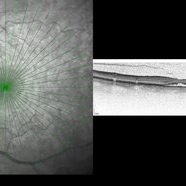
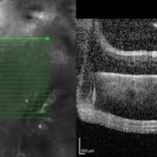
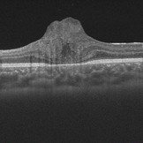
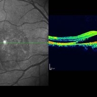
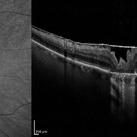
 3D OCT of juxtapapillary melanoma
May 15 2020 by Sophia El Hamichi, MD
A 63-year-old male with juxtapapillary melanoma of the right eye. Visual acuity at presentation was 20/25 OD. Patient treated with brachytherapy Iodine125 plaque
Photographer: Belinda Rodriguez
Condition/keywords: optical coherence tomography (OCT)
-
 5 Weeks Post JETREA
May 15 2013 by Robert T. Wendel, MD
5 weeks post-op JETREA for stage 2 MH. OCT shows closing macular hole, but the cortex still attached.
Condition/keywords: macular hole, optical coherence tomography (OCT)
-
 Active CNVM
Jul 11 2016 by Manish Nagpal, MD, FRCS (UK), FASRS
Colour photo showing an active CNVM.
Photographer: pooja barot
Condition/keywords: choroidal neovascular membrane (CNVM), optical coherence tomography (OCT)
-
 Active CNVM on Angio OCT
Jul 11 2016 by Manish Nagpal, MD, FRCS (UK), FASRS
Angio OCT picture showing neovascularization corresponding to the area of CNVM.
Photographer: pooja barot
Condition/keywords: choroidal neovascular membrane (CNVM), optical coherence tomography (OCT)
-
 Advanced PDR
Sep 1 2014 by Hamid Ahmadieh, MD
OCT image of the right eye of a 50-year-old woman with advanced PDR.
Photographer: Soodabeh Fooladian, Negah Eye Center, Tehran, Iran
Condition/keywords: optical coherence tomography (OCT), proliferative diabetic retinopathy (PDR)
-
 Advanced PDR
Sep 1 2014 by Hamid Ahmadieh, MD
OCT image of the left eye of a 50-year-old woman with advanced PDR.
Photographer: Soodabeh Fooladian, Negah Eye Center, Tehran, Iran
Condition/keywords: optical coherence tomography (OCT), proliferative diabetic retinopathy (PDR)
-
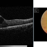
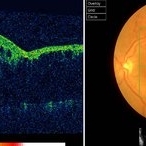
 Aneurysm OCT
Sep 17 2019 by Zachary M Bodnar, MD
Aneurysm OCT
Condition/keywords: optical coherence tomography (OCT), retinal arterial macroaneurysm
-
 Angioid Streaks & CNV (Fig 5)
Sep 2 2012 by Hamid Ahmadieh, MD
OCT imaging of a 53-year-old woman with a juxtafoveal CNV secondary to angioid streaks.
Photographer: Hamid Ahmadieh, Ophthalmic Research Center, Labbafinejad Medical Center
Imaging device: Topcon
Condition/keywords: angioid streaks, choroidal neovascularization (CNV), optical coherence tomography (OCT)
-
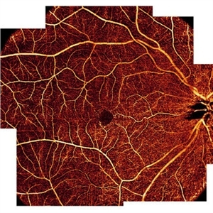 AngioOCT Normal Widefield Scan
May 8 2015 by Timur Shaimov
Optical coherence tomography angiography of a 28-year-old woman without any macular pathology. Seven 6x6mm angioOCT EnFace images used to merge into widefield view. We used the Optovue RTVue XR Avanti (Optovue, USA) optical coherence tomography system with split-spectrum amplitude decorrelation angiography algorithm (SSADA).
Photographer: Timur Shaimov
Imaging device: Optovue RTVue XR Avanti
Condition/keywords: optical coherence tomography (OCT)
-
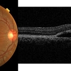
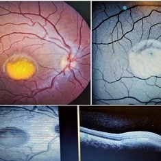
 Astrocytoma OCT
Jan 9 2018 by Sidra Zafar
Swept source OCT imaging of retinal astrocytoma in a female child with known diagnosis of tuberous sclerosis.
Imaging device: Swept Source
Condition/keywords: astrocytoma, optical coherence tomography (OCT), tuberous sclerosis
-
 Autosomal Dominant Drusen of Bruch's Membrane, OCT
May 3 2018 by Alexandr Stepanov
Autosomal dominant drusen of Bruch's membrane, OCT.
Photographer: Alexandr Stepanov MD, PhD, FEBO, Faculty Hospital Hradec Kralove, Czech Republic
Condition/keywords: Bruch's membrane, drusen, optical coherence tomography (OCT)
-
 AZOOR OCT 10-10-13
Dec 14 2013 by Robert T. Wendel, MD
AZOOR OCT 10-10-13
Condition/keywords: acute zonal occult outer retinopathy (AZOOR), optical coherence tomography (OCT)
-
 AZOOR OCT2 10-10-13
Dec 14 2013 by Robert T. Wendel, MD
AZOOR OCT2 10-10-13
Condition/keywords: acute zonal occult outer retinopathy (AZOOR), optical coherence tomography (OCT)
-
 Behcet's Disease
Mar 13 2013 by Hamid Ahmadieh, MD
OCT of the right eye of a 23-year-old man with retinal vasculitis and branch retinal vein occlusion (BRVO) due to Behcet's disease .
Photographer: Solmaz Shahmohammad, Negah Eye Center, Tehran
Imaging device: Topcon OCT
Condition/keywords: branch retinal vein occlusion (BRVO), optical coherence tomography (OCT), retinal vasculitis
-
 Best Disease
Oct 10 2015 by Hamid Ahmadieh, MD
Color fundus photograph and OCT image of the right eye of a 35-year-old man with decreased vision due to Best disease.
Photographer: Shabnam Pooreh, Negah Eye Center, Tehran, Iran
Condition/keywords: Best disease, color fundus photograph, optical coherence tomography (OCT), vitelliform lesion
-
 Best Disease
Oct 10 2015 by Hamid Ahmadieh, MD
Color fundus photograph and OCT image of the left eye of a 35-year-old man with decreased vision due to Best disease.
Photographer: Shabnam Pooreh, Negah Eye Center, Tehran, Iran
Condition/keywords: Best disease, color fundus photograph, optical coherence tomography (OCT), vitelliform lesion
-
 Best Disease
Mar 9 2013 by Hamid Ahmadieh, MD
OCT of the right eye of a 49-year-old man with decreased VA due to advanced Best disease.
Photographer: Soodabeh Fooladin, Negah Eye Center, Tehran
Imaging device: Heidelberg Spectralis
Condition/keywords: Best disease, optical coherence tomography (OCT)
-
 Best Disease
Mar 9 2013 by Hamid Ahmadieh, MD
OCT of the left eye of a 49-year-old man with decreased VA due to advanced Best disease.
Photographer: Soodabeh Fooladin, Negah Eye Center, Tehran
Imaging device: Heidelberg Spectralis
Condition/keywords: Best disease, optical coherence tomography (OCT)
-
 Best Vitelliform Macular Dystrophy
Mar 17 2020 by Sophia El Hamichi, MD
Classic "egg yolk" presentation in a 16-year-old female with best disease.
Condition/keywords: autofluorescence imaging, Best disease, optical coherence tomography (OCT), vitelliform macular dystrophy
-
 Biclonal Gammopathy
Jan 29 2014 by Mallika Goyal, MD
OCT of the left eye of a 17-year-old girl with monoclonal gammopathy shows extreme thinning of the foveal centre, likely from ischaemia associated atrophy.
Photographer: Mallika Goyal, MD, Apollo Health City, Hyderabad, India
Condition/keywords: biclonal gammopathy, optical coherence tomography (OCT)
-
 Biclonal Gammopathy
Jan 29 2014 by Mallika Goyal, MD
OCT of the right eye of a 17-year-old girl with biclonal gammopathy shows extreme macular elevation secondary to a thick epimacular membrane.
Photographer: Mallika Goyal, MD, Apollo Health City, Hyderabad, India
Condition/keywords: biclonal gammopathy, optical coherence tomography (OCT)
-
 Branch Retinal Vein Occlusion (BRVO)
Sep 11 2012 by Hamid Ahmadieh, MD
Color fundus photograph and OCT image of a 60-year-old woman with a recent onset BRVO.
Photographer: Hamid Ahmadieh, MD, Ophthalmic Research Center, Labbafinejad Medical Center, Shahid Beheshti University of Medical Sciences
Imaging device: Topcon
Condition/keywords: branch retinal vein occlusion (BRVO), optical coherence tomography (OCT)
-
 Bulls eye retinopathy
Nov 20 2012 by Roy Schwartz, MD
75-YEAR-OLD FEMALE PRESENTS WITH BILATERAL GRADUAL VISUAL LOSS 6/30 Dx BE PSEUDOPHAKIA + PCO BE BULLS EYE MACULOPATHY PER FA VA IMPROVES TO 6/10 S/P YAG CAPSULOTOMY OCT - BE MACULAR SUBRETINAL FLUID NO HISTORY OF CHLOROQUINE THERAPY NO DRUSEN OR SIGNS OF AMD WORKING DIAGNOSIS - BE CHRONIC CSCR
Imaging device: Heidelberg spectralis
Condition/keywords: bull's eye maculopathy, optical coherence tomography (OCT)
-
 Bulls eye retinopathy OCT LE
Nov 20 2012 by Roy Schwartz, MD
75-YEAR-OLD FEMALE PRESENTS WITH BILATERAL GRADUAL VISUAL LOSS 6/30 Dx BE PSEUDOPHAKIA + PCO BE BULLS EYE MACULOPATHY PER FA VA IMPROVES TO 6/10 S/P YAG CAPSULOTOMY OCT - BE MACULAR SUBRETINAL FLUID NO HISTORY OF CHLOROQUINE THERAPY NO DRUSEN OR SIGNS OF AMD WORKING DIAGNOSIS - BE CHRONIC CSCR
Imaging device: Heidelberg spectralis
Condition/keywords: bull's eye maculopathy, chronic central serous chorioretinopathy (CSCR), optical coherence tomography (OCT)
-
 Bullseye Maculopathy
Jan 22 2024 by Kali Jend
Optical coherence tomography of a 73-year-old female with Bullseye Macular Changes affecting her left eye. Patient reports having a family history of this condition and denies prior Plaquenil or Elmiron use. Compared to previous imaging, the patient's condition progressed in the left eye from 2020 to 2023. Patient has a history of fluctuating Diabetic Macular Edema and a current Epiretinal Membrane as well. Patient's vision was Ncc20/60 at the time the image was taken.
Photographer: Kali Jend
Imaging device: Heidelberg Spectralis
Condition/keywords: bullseye maculopathy, epiretinal membrane (ERM), heidelberg spectralis, left eye, macular pucker, OCT, optical coherence tomography (OCT)
 Loading…